Sunday, November 27 | 12:05 p.m.-12:15 p.m. | SSA15-09 | Room N229
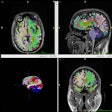
Researchers from Ohio State University (OSU) Medical Center have concluded that a combination of MRI techniques -- diffusion-tensor imaging (DTI) and susceptibility-weighted imaging -- at a 7-tesla field strength could improve the clinical diagnosis of chronic mild traumatic brain injury and provide additional information on underlying tissue microstructure.Seven mild traumatic brain injury patients with long-term persistent symptoms were evaluated with six approximately age-matched healthy controls. All 13 subjects received susceptibility-weighted imaging and DTI at 7 tesla.
Images were reconstructed using high-pass filtering of complex susceptibility-weighted imaging data, and fractional anisotropy and higher apparent diffusion coefficient maps were computed for DTI.
The researchers found that 7-tesla DTI results were comparable to previously published findings in chronic mild traumatic brain injury, while susceptibility-weighted imaging phase and magnitude images showed differences. The results also indicate that susceptibility-weighted imaging can differentiate between mild traumatic brain injury and control groups.
While the findings are preliminary, presenter Sharon Schreiber, a graduate research associate at OSU Medical Center, said clinical benefits are a "more neuroanatomically relevant classification of traumatic brain injury, which could be used to form more accurate diagnoses and prognoses and help guide the development and use of pharmaceutical therapies to halt further damage associated with pathology."
Schreiber and colleagues plan to continue their research, given the encouraging early results.
"The results show promise for investigating a more quantitative approach more thoroughly, which includes assessing the effect of image postprocessing techniques on the phase parameters which correlate to microstructure, as well as truly combining information gained from both diffusion-tensor imaging and susceptibility-weighted imaging to give the most sensitive and accurate classifier," she added.


.fFmgij6Hin.png?auto=compress%2Cformat&fit=crop&h=100&q=70&w=100)



.fFmgij6Hin.png?auto=compress%2Cformat&fit=crop&h=167&q=70&w=250)


