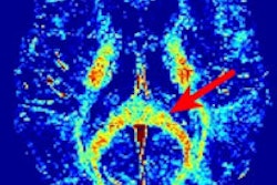
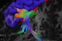
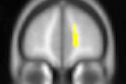
Using MRI with a diffusion-tensor imaging (DTI) protocol, researchers have found that the brain scans of high school football and hockey players who have taken routine hits to the head during play show subtle injury, even when there is no evidence of a concussion, according to a study published online in Magnetic Resonance Imaging.
Lead study author Dr. Jeffrey Bazarian, associate professor of emergency medicine at the University of Rochester Medical Center, and colleagues applied a new statistical approach to diffusion-tensor imaging to detect white-matter changes in the teenage athletes' brains.
Nine athletes and six people in a control group volunteered to participate in the study during the 2006-2007 sports season. Among the nine athletes, one was diagnosed with a sports-related concussion that season, but six others sustained many subconcussive blows. The brains of those six athletes showed abnormalities on postseason scans that more closely resembled the concussed brain than those in the control group.
Many changes were found in the brain of the player with the diagnosed concussion, according to the researchers. However, an intermediate level of change also occurred among the players who reported from 26 to 399 total subconcussive blows. The fewest changes occurred in the control group.


