Researchers at the University of California, San Francisco (UCSF) have used MRI to reveal a molecular marker in tissue samples from brain tumors that has been linked to better survival.
By monitoring this marker in the brain, doctors may have a better way to follow patients after surgery, according to an article published in the January issue of Science Translational Medicine.
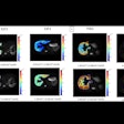
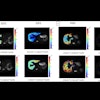
The study used MRI to obtain data from image-guided tissue samples from more than 50 patients with gliomas. The samples demonstrated the presence of a chemical called 2-HG, which is linked to mutations in a gene known as IDH1.
Recent studies have shown that these mutations are more common in low-grade tumors and are associated with longer survival. More than 70% of patients with low-grade gliomas have mutations to the IDH1 genes in their cancer cells.
Study co-author Sarah Nelson, PhD, a professor in UCSF's department of radiology and biomedical imaging, said the techniques must be refined for standard hospital MRI scanners to image the presence of 2-HG.

.fFmgij6Hin.png?auto=compress%2Cformat&fit=crop&h=100&q=70&w=100)
.fFmgij6Hin.png?auto=compress%2Cformat&fit=crop&h=167&q=70&w=250)
