Tuesday, November 27 | 3:40 p.m.-3:50 p.m. | SSJ04-05 | Room S504AB
Researchers from Leipzig Heart Center in Germany plan to discuss what they say is the first pilot electrophysiology (EP) study of real-time MRI-guided placement of multiple catheters in humans with subsequent performance of stimulation maneuvers.The study included four men and one woman (mean age, 64.4 years) with symptomatic arrhythmias. Three people with typical atrial flutter presented for isthmus ablation, one person presented for an EP study, and the remaining person presented for slow pathway ablation for atrioventricular nodal re-entrant tachycardia.
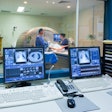
After the procedures, all five patients were transferred to a 1.5-tesla whole-body MRI system (Intera, Philips Healthcare) for an EP diagnostic procedure. Two MRI-compatible diagnostic/ablation catheters were inserted through femoral sheaths and manipulated by an experienced electrophysiologist using a commercially available, interactive, real-time steady-state free-precession MRI sequence.
Using passive catheter tracking, all catheters were placed successfully in the right ventricle and the right atrium. The placement was confirmed by intracardiac electrograms. Furthermore, simple programmed stimulation maneuvers were performed. During and after the procedure, no adverse effects were observed in any patients.
"Interventional procedures in rhythmology, such as electrophysiology and ablation, can be rather time-consuming due to corresponding long fluoroscopy times and can involve high radiation exposure for the patient and the interventionist," said lead study author Dr. Matthias Gutberlet, PhD, chair of the department of diagnostic and interventional radiology. "MRI helps to reduce exposure for the patients and staff, at least in the present transition period in which a fluoroscopy or x-ray backup is necessary."
With MRI techniques already in routine use for visualizing acute infarction or inflammation, it may also be possible to assess atrial tissue properties prior to, during, and after radiofrequency ablation, Gutberlet noted. This technique can be highly relevant to an electrophysiologist when treating patients with complex arrhythmias and may constitute a step toward more personalized treatment in rhythmology.
"We are not totally sure as yet about the added value for MRI-guided ablations," Gutberlet added. "Our first experiences with MR-guided electrophysiology procedures and published data from other groups in the area of image-guided therapy led to the following hypothesis: Being able to see the catheter within the real cardiac anatomy ... may indeed facilitate interventional procedures."
Gutberlet and colleagues plan to continue their research; ablations of atrial flutter have already been performed in the MRI system and will be continued with a modified catheter. "Furthermore, besides passive catheter tracking, active tracking is also on our road map," he said.


.fFmgij6Hin.png?auto=compress%2Cformat&fit=crop&h=100&q=70&w=100)


.fFmgij6Hin.png?auto=compress%2Cformat&fit=crop&h=167&q=70&w=250)


