Monday, December 2 | 10:40 a.m.-10:50 a.m. | SSC02-02 | Room S502AB
German researchers used MR images to show how a relatively short period of high-intensity training in men can greatly influence left and right ventricular characteristics and function.Lead author Dr. Michael Scharf and colleagues from University Hospital Erlangen concluded that these findings are not associated with pathologic features predisposing individuals to sudden cardiac death.
The researchers randomly assigned 42 untrained volunteers, ranging in age from 33 to 51 years, to a high-intensity training group, while another 42 individuals in the same age span were assigned to an inactive control group.
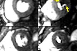
The participants underwent cardiac MRI to assess myocardial morphology and function of the left and right ventricle before and after four months, during which the high-intensity group underwent training.
Subjects also took part in a progressive-intensity treadmill test that assessed ventilation parameters and heart rate at the anaerobic threshold. Ejection fraction, end-diastolic volume, end-systolic volume, stroke volume, myocardial mass, and cardiac index were measured for the left and right ventricle.
After training, indexed volume and mass for the left and right ventricles were "significantly greater" in the high-intensity group than in the inactive control group, which remained unchanged, according to the researchers.

.fFmgij6Hin.png?auto=compress%2Cformat&fit=crop&h=100&q=70&w=100)




.fFmgij6Hin.png?auto=compress%2Cformat&fit=crop&h=167&q=70&w=250)
