Monday, December 2 | 11:20 a.m.-11:30 a.m. | SSC12-06 | Room N229
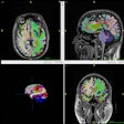
Researchers at NYU Langone Medical Center have found a connection between levels of thalamic iron and microstructural changes in the brain's frontal white matter in patients with mild traumatic brain injury (MTBI).The group used 3-tesla diffusion-tensor MRI (DTI-MRI) to assess frontal white-matter microstructure changes and magnetic field correlation (MFC) to measure thalamic iron. The findings suggest that iron may contribute to secondary injury after mild TBI.
Complications from mild TBI are difficult to detect using conventional CT and MRI, said study co-author Dr. Yvonne Lui, chief of the neuroradiology section at NYU Langone. "Current research uses novel techniques to try to identify [these] abnormalities."
Previous research has found that thalamic iron increases after a concussion, and in MTBI, there is particular interest in frontocortical connections to areas that control executive function.
Lui and colleagues prospectively enrolled 27 patients with confirmed MTBI; longitudinal data were available for 14 subjects. Subjects underwent 3-tesla DTI-MRI and microscopic magnetic field correlation one month and one year after their injury.
One year after injury, there was a link between higher thalamic iron measures and frontal white-matter microstructural changes. The study is the first to report a connection between white-matter injury and iron accumulation in MTBI patients, Lui and colleagues noted.
"We still know little about the pathophysiology of disease after mild TBI," Lui said. "Trying to understand the underlying mechanisms of disease will allow for early stratification of patients and pave the way to targeted therapy."


.fFmgij6Hin.png?auto=compress%2Cformat&fit=crop&h=100&q=70&w=100)


.fFmgij6Hin.png?auto=compress%2Cformat&fit=crop&h=167&q=70&w=250)


