Monday, December 2 | 8:50 a.m.-9:00 a.m. | VSBR21-02 | Arie Crown Theater
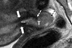
7-tesla MRI matches or surpasses 3-tesla imaging in terms of quality and uniformity in breast imaging, according to researchers from NYU Langone Medical Center.In this Monday morning scientific session, Dr. Linda Moy and colleagues will discuss findings from a study they conducted with 17 women using T1-weighted fat-suppressed breast MRI at 7 tesla. The group used 3-tesla images in the same patients as a baseline reference.
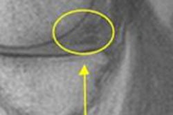
Two radiologists qualitatively graded the images on a five-point scale and quantitatively assessed them for fibroglandular/fat contrast and signal uniformity. Moy's group found that in standard resolution images the quality scores at 7 tesla and 3 tesla were similar (4.3 versus 4.1, respectively). But 7-tesla MRI had a better signal-to-noise ratio than 3-tesla MRI in high-resolution images (4.2 versus 3.1, respectively), which allowed clinicians to see ligaments and dendritic patterns more clearly.
Clinicians could make use of this improved signal-to-noise ratio for high-resolution imaging to improve fibroglandular tissue detail and classification of lesions, the researchers concluded.


