CHICAGO - An MRI technique called magnetic field correlation is being used to measure iron levels in the brains of children with attention deficit/hyperactivity disorder (ADHD) to improve ADHD diagnosis, according to a study presented at RSNA 2013.
While the results are preliminary, the researchers from the Medical University of South Carolina (MUSC) are hopeful that MFC could eventually help physicians and parents make more informed decisions about treatment for children and adolescents with ADHD.
MFC was developed by study co-authors and MUSC faculty members Joseph Helpern, PhD, and Jens Jensen, PhD, and is designed to measure iron levels through a decrease in MRI signal. Simply put, as the level of iron increases, there is a corresponding decrease in the MRI signal, which creates dark spots on the image.
 Vitria Adisetiyo, PhD, from MUSC.
"You can actually see areas where we know there is a high level of iron, such as the basal ganglia region; you will see a drop in the MFC," lead author Vitria Adisetiyo, PhD, a postdoctoral research fellow in MUSC's Center for Biomedical Imaging, told AuntMinnie.com.
Abnormal iron metabolism in the brain has been associated with neurodegenerative disorders. Previous research has found that children with iron deficiency in the brain develop slower, have issues with cognition and reduced attention span, and can be lethargic. Children with ADHD often are hyperactive, do not stay focused, and find it difficult to control their behavior.
ADHD treatment
Psychostimulant medications such as Ritalin are commonly given to children and adolescents to reduce ADHD symptoms. The drugs are designed to regulate levels of the neurotransmitter dopamine.
Because iron is required for dopamine synthesis, assessing iron levels with MRI may provide a noninvasive, indirect measure of dopamine.
"The reason we were looking at brain iron was because there is a small but growing amount of literature that identifies some type of iron homeostasis abnormality in the disorder," Adisetiyo said. "Given the fact that we had tools that could be more sensitive and specific to measuring iron, we thought, 'Let's see if we can use this new tool to further investigate this finding.' "
The researchers measured brain iron in 22 children and adolescents with ADHD and 27 healthy controls. Subjects ranged in age from 8 to 18 years. MRI was conducted on a 3-tesla scanner (Magnetom Trio, Siemens Healthcare) with no contrast agent.
"The technique uses endogenous iron as the imaging agent, so there is nothing injected," Adisetiyo added.
The results showed that 12 ADHD patients who had never been on medication had significantly lower iron levels than the 10 ADHD patients who had been on psychostimulant medication and the 27 controls. The lower brain iron levels among nonmedicated subjects appeared to normalize with psychostimulant medication.
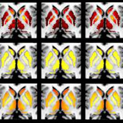
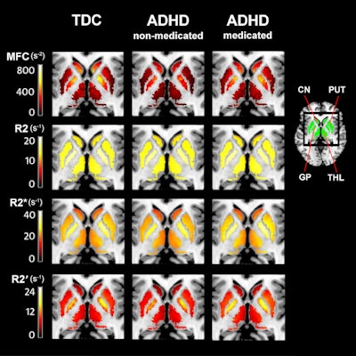
 Images show nonmedicated ADHD patients have reduced striatal and thalamic MFC measures of brain iron, compared with controls and medicated ADHD patients. Brain iron measures in the putamen (PUT), caudate nucleus (CN), thalamic (THL), or globus pallidus (GP) did not differ between controls and medicated ADHD patients. The statistically significant differences are visible in the MFC group maps (top row) but not in conventional relaxation rate maps in the second, third, and bottom rows. Image courtesy of RSNA and Vitria Adisetiyo, PhD.
While the study is preliminary, Adisetiyo said the results are exciting and validate the continuation of the research with a larger cohort.
"Once we are able to replicate the results in a bigger study and are able to distinguish the groups more clearly, then we can talk about implementing it in the clinic," she said. "But at this point I would be cautious about calling it a diagnostic tool."
The hope is that MFC could be helpful when, for example, a psychiatrist is not fully confident of a diagnosis based on traditional methods. In addition, MFC is noninvasive and does not use contrast. Currently, dopamine levels in children and adolescents with ADHD are measured using radioactive contrast agents, which most parents often decline for their children.
Follow-up research
Adisetiyo and colleagues are currently scanning more subjects using MFC to expand their research.
"One of the limitations of the current dataset is that it is cross-sectional, meaning that we could not follow a subject or participant before and after medication treatment," she said. "We sampled children who were either naive of medication or kids who were already on medication."
Therefore, the next step is to conduct a longitudinal study with larger cohorts to follow children both before and after psychostimulant medication to see if their brain iron levels are initially low and then increase with medication, as was seen in this study.
The MUSC researchers also are pursuing a slightly more controversial avenue that touches upon the issue of addiction in ADHD. Psychostimulant medications can become addictive in some patients and lead to abuse of other psychostimulant substances such as cocaine.
"What we hope to look at is brain iron levels in cocaine addicts," Adisetiyo said. "It would be a completely separate study, but it would also assess the range of iron levels in people who we suspect have low iron levels, which is kids with ADHD. Normal people have regular iron levels and cocaine addicts we assume have higher iron levels because they are bombarding their system with psychostimulants."


