Resting-state functional MRI (fMRI) has uncovered new abnormalities in key brain regions in children and adolescents with attention-deficit/hyperactivity disorder (ADHD), according to a study from China published online April 30 in Radiology.
Researchers from West China Hospital at Sichuan University discovered that children with ADHD have altered structure and function in brain regions involved in emotion control and strategic planning.
The findings not only indicate more widespread brain alterations in ADHD patients than previously seen, they show the potential of resting-state fMRI for providing an accurate and early diagnosis, according to the authors. That information, in turn, could help physicians monitor ADHD progression and provide more appropriate clinical treatment (Radiology, April 30, 2014).
Unlocking the mystery
Resting-state fMRI is a relatively new technique designed to assess neural function when the brain is not focused on a specific task. It can be used to determine the brain's functional organization independent of task performance.
While previous fMRI studies have explored the brain's frontostriatal circuit, which consists of neural pathways that help to control behavior and attention, the specific brain physiology underlying ADHD remains somewhat of a mystery, noted lead author Fei Li, PhD, from Huaxi MR Research Center, and colleagues.
The study included 33 boys (mean age, 10.1 ± 2.6 years) with ADHD who were not receiving medication and had no comorbidities, along with 32 healthy control subjects (mean age, 10.9 ± 2.6 years). The subjects were recruited and studied between June 2009 and December 2011 at the Mental Health Center at West China Hospital.
All participants underwent resting-state fMRI in a 3-tesla scanner (Magnetom Trio, Siemens Healthcare) with an 8-channel phased-array head coil.
Regional brain activity was measured using amplitude of low-frequency fluctuation (ALFF), while brain function was evaluated by measuring functional connectivity. The calculations were later compared between ADHD patients and the control subjects.
The researchers also assessed the participants' behavioral performance with a series of tests for executive function, which included their ability to plan, organize, manage time, and control emotions. Performances were scored using the Wisconsin Card Sorting Test (WCST) and the Stroop color-word test.
Brain abnormalities
In reviewing the resting-state fMR images, Li and colleagues found significant ALFF abnormalities in five brain regions in the ADHD patients compared with the control subjects.
More specifically, patients with ADHD showed impaired executive function, along with the following:
- Lower ALFF in the left orbitofrontal cortex and the left ventral superior frontal gyrus
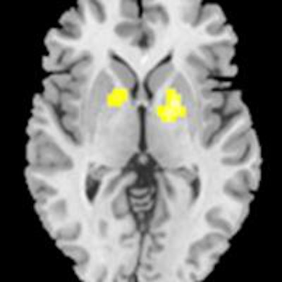
- Higher ALFF in the left globus pallidus, the right globus pallidus, and the right dorsal superior frontal gyrus
- Lower long-range functional connectivity in the frontoparietal and frontocerebellar networks
- Higher functional connectivity in the frontostriatal circuit that correlated across subjects with ADHD with the degree of executive dysfunction
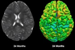
 Images show differences in ALFF between ADHD and control subjects. Regions of increased ALFF in ADHD subjects are indicated by red, orange, and yellow; decreased ALFF areas are depicted in white, light blue, and dark blue. Images courtesy of Radiology.
Subjects with ADHD performed much worse than control subjects on the study's executive function tests, the authors wrote. The ADHD patients had fewer total correct numbers and completed fewer categories on the WCST, and they committed more total errors than their control counterparts.
In the Stroop color-word test, the ADHD patients had fewer correct numbers and more errors, and they took longer to complete the test than control subjects.
"Our study suggests that the structural and functional abnormalities in these brain regions might cause the inattention and hyperactivity of the patients with ADHD, and we are doing further analysis on their correlation with the clinical symptoms," said senior author Dr. Qiyong Gong, PhD, in an RSNA release.
Larger studies are needed to validate the results, he said. The researchers also plan to study changes in connectivity over time in ADHD patients and explore the potential differences of functional connectivity between the clinical subtypes of ADHD, such as inattentiveness and hyperactivity.


