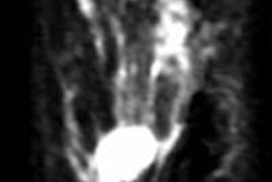
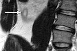
A new breast-conserving surgical technique combines presurgical MRI with optical scanning during surgery to locate small breast tumors for excision, according to a pilot study published online in March in the Annals of Surgical Oncology.
Physicians from Dartmouth-Hitchcock Norris Cotton Cancer Center collaborated with engineers from Dartmouth's Thayer School of Engineering to develop the technique, which gives surgeons 3D images of breast cancer during surgery, said senior author Dr. Richard Barth Jr. in a statement.
Traditionally, a wire has been inserted into the breast to mark small tumors before surgery, but this technique isn't very accurate, leaving cancer cells behind in 30% to 40% of cases. The new technique uses preoperative MRI for tumor mapping and an optical scan to identify the tumor's size, shape, and location; it then combines the two to create a 3D image for the surgeon.
The pilot study was funded by the Dartmouth Clinical and Translational Science Institute.


