Canadian researchers have developed a new MRI technique to better track the progression of multiple sclerosis (MS).
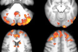
Called quantitative susceptibility (QS) MRI, the technique measures damage in specific areas of the brain, and the study showed this damage to be common to all subjects. Results were published online May 14 in Radiology.
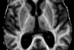
Senior author Ravi Menon, PhD, from the University of Western Ontario, said that conventional MRI scans of multiple sclerosis patients show lesions in the brain very clearly, but the number or volume of the lesions do not correlate with the patients' disabilities. Clinicians have been aware of the paradox since the early 1980s, but MRI remains the only imaging tool available to assess the disease.
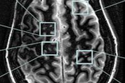
QS MRI shows considerable damage occurring in common areas in both the white and gray matter. These QS measures correlate with disease symptoms, Menon said. The process uses a standard 3-tesla MRI scanner, so it could be reproduced in any hospital.
The researchers mapped QS in 25 patients with relapsing-remitting multiple sclerosis or clinically isolated syndrome. The study also included 15 age- and sex-matched control subjects.
QS MRI showed common areas of damage in all patients. The areas correlated well with a test to measure multiple sclerosis progression, as well as with age and time since diagnosis, the group concluded.


