Joint cracking in the fingers appears to be caused by the rapid formation of a gas-filled cavity in the joints' synovial fluid, rather than the collapse of pre-existing bubbles of air as the joints separate, according to a new study in PLOS One that used static and real-time MRI.
Canadian and Australian researchers used cine MRI to visualize joint cracking in real-time -- a technology that has not been used before for this purpose, according to lead author Greg Kawchuk, PhD, of the University of Alberta in Edmonton.
 Greg Kawchuk, PhD, from the University of Alberta.
"We realized there was a decades-old debate in the literature as to what causes joint cracking, and we also realized that up until now, nobody had actually observed the events of joint cracking in sufficient detail, in real-time," Kawchuk told AuntMinnie.com via email. "Given that, we decided to conduct the study with the hope of settling the debate by better understanding the mechanisms underlying joint cracking."
Kawchuk's group used a Magnetom Sonata (Siemens Healthcare) 1.5-tesla MRI system with a finger coil provided by the company. For the study, the single participant lay prone with his hand attached to the radiofrequency coil, which was then threaded through the magnet. The team took static images of the subject's 10 metacarpophalangeal (MCP) joints before and after taking a cine MR acquisition, during which force was increasingly applied manually through the coil until the study participant indicated that the joint had cracked (PLOS One, April 15, 2015).
The images were reviewed by an imaging physicist and two radiologists, the authors wrote.
"MRI suites are not set up for this kind of study, so we had to make some special equipment as well as devise some unique strategies to observe joint cracking within the magnet," Kawchuk told AuntMinnie.com.
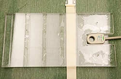
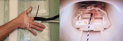 The radiofrequency coil inside the clear housing (top). The MCP joint of interest centered over the bore of the
radiofrequency coil (above left). The participant's hand within the imaging magnet (above right). All images courtesy of PLOS One.
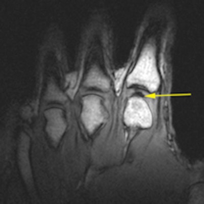
All 10 of the subject's MCP joints cracked. The static MR images showed normal MCP joints without any gas bubbles before cracking; after cracking, static imaging showed a dark intra-articular space, according to Kawchuk's team.
The study suggests that joint cracking is caused by tribonucleation, a process by which gas bubbles are created when solid surfaces make or break contact in liquid that contains dissolved gas.
"Our results offer direct experimental evidence that joint cracking is the result of cavity inception within synovial fluid rather than a collapse of a pre-existing bubble," the group wrote.
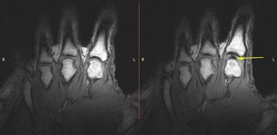 T1 static images of the hand in the resting phase before cracking (left). The same hand following cracking with the addition of a postcracking distraction force (right). Note the dark void (yellow arrow).
A surprising finding was that the researchers observed a white flash just before the joint cracked, Kawchuk said.
"The white signal we saw in the joint just before it cracked was likely caused by fluid collecting between the joint surfaces as tension increased," he said. "We'd like to explore if this is a sign of cartilage health, which could offer a noninvasive way of determining a patient's joint status."
Kawchuk and colleagues hope that by defining the process underlying joint cracking, its therapeutic benefits -- or possible harms -- might be better understood.
"The ability to visualize this phenomenon could allow us to better understand the resiliency of joints and why some joints lose that resiliency," he said.


