Dear AuntMinnie Member,
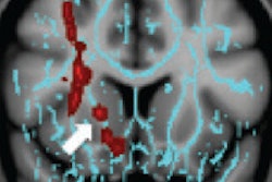
A new animal study on gadolinium-based contrast agents for MRI scanning found persistent abnormalities in the brains of rats that received a leading agent.
French researchers used MRI to track brain changes in rats that were given gadodiamide, comparing them with other rats that received gadoterate meglumine or a saline solution. They found much higher gadolinium concentrations after scanning in the gadodiamide group than in the rats that received gadoterate meglumine.
The findings are concerning given other recent studies that indicate that gadolinium can persist in the brain years after MRI contrast is administered. They also reinforce suspicions that the severity of reactions to gadolinium could vary depending on the type of agent being used.
But the current study compared only two gadolinium contrast agents, the research involved animals rather than humans, and, what's more, the lead author on the study was a scientist with the company that makes gadoterate meglumine, which was found to have lower persistence in the brain.
Still, should we be concerned? Learn more by clicking here, or visit our MRI Community at mri.auntminnie.com.
CT radiation surprise
Another study raising concerns about the safety of medical imaging was published this week in JAMA Internal Medicine by California researchers, who found wide variation in the levels of radiation used in CT scans of individuals suspected of having kidney stones.
The researchers were actually trying to assess how initial imaging with CT compared to ultrasound for individuals who might have urolithiasis. But a funny thing happened on the way to data analysis: Radiation dose levels for the CT exams were all over the board, and most were way too high.
The researchers were surprised by the variance and expressed regret that they hadn't established unified scanning protocols in the design of the trial. They believe the results show that voluntary efforts to monitor and control radiation dose are not enough.
Read more by clicking here, or visit our CT Community at ct.auntminnie.com.
Hybrid 3D printing
Finally, visit our Advanced Visualization Community for a fascinating article on a breakthrough in 3D printing from medical imaging scans.
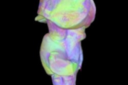
A group from Michigan has created hybrid 3D printed models of the heart using not one but two medical imaging modalities: CT and echocardiography.
They believe the advance could lead to more accurate and realistic 3D models, as each imaging modality has certain strengths that contribute to the reconstructions. Read more by clicking here, or visit the community at av.auntminnie.com.


.fFmgij6Hin.png?auto=compress%2Cformat&fit=crop&h=100&q=70&w=100)


.fFmgij6Hin.png?auto=compress%2Cformat&fit=crop&h=167&q=70&w=250)


