Dear MRI Insider,
Kids with heart abnormalities often present complex problems for clinicians and surgeons, given that few cases are alike. Pediatric heart surgeons therefore require as much visual information as possible to correct an abnormality.
To help in those situations, researchers from the Massachusetts Institute of Technology and Boston Children's Hospital have developed an automated software method to convert 3D MRI scans of a patient's heart into a tangible model that surgeons can use to plan surgery.
While the endeavor to create heart models from 3D MRI scans is not unique, this initiative is looking to craft a faster and more automated process to define the borders and contours of pediatric hearts and facilitate the production of 3D heart models. As an Insider subscriber, you have an exclusive first look at how the venture is progressing.
In other news, a study published online today in JAMA Oncology questions whether preoperative breast MRI really helps patients. Researchers from Canada found that MRI scans before breast surgery led to higher rates of mastectomy and biopsy and longer delays between diagnosis and surgery. However, they didn't measure long-term outcomes such as survival rates. Read more by clicking here.
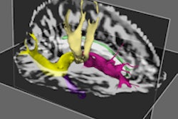
Meanwhile, Italian researchers using diffusion-tensor MRI (DTI-MRI) have found that high blood pressure can damage nerve tracts that connect different parts of the brain. They used the technique to reconstruct white-matter connections that correlate with specific cognitive functions to determine if damage to the connections might be a precursor to the development of dementia through vascular disease.
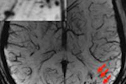
The sooner a person with a traumatic brain injury (TBI) undergoes an MRI scan, the better the chances of finding microbleeds in the brain and beginning treatment for the abnormality. That conclusion comes from a study of military personnel in which approximately one-quarter of the patients had evidence of cerebral microhemorrhages within three months of their injury. However, the number of detected bleeds decreased dramatically in the months afterward.
While some traditions should be valued and retained, the old standard free-text radiology report might not be one of them. Brigham and Women's Hospital is using a structured reporting template to achieve a significant improvement in the quality of MRI reports for the crucial task of staging rectal cancer.
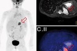
Finally, despite its slightly longer scan time, young pediatric patients may benefit from FDG-PET/MRI for staging and clinically evaluating oncologic disorders, given the hybrid modality's superior soft-tissue contrast and significantly lower radiation dose, concluded researchers from Germany.
Be sure to stay in touch with the MRI Community on a daily basis as we cover groundbreaking research from around the world and its contribution to clinical practices.


.fFmgij6Hin.png?auto=compress%2Cformat&fit=crop&h=100&q=70&w=100)


.fFmgij6Hin.png?auto=compress%2Cformat&fit=crop&h=167&q=70&w=250)


