Dear AuntMinnie Member,
Shear-wave elastography could be a better tool than other ultrasound modalities for detecting tendinopathy and assessing response to treatment.
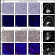
That's according to a new study in our Ultrasound Community by German researchers, who compared shear-wave elastography with B-mode and power Doppler for diagnosing tendinopathy. The technique's measurement of tissue stiffness makes it a good tool for tendinopathy, because diseased tendons are softer than normal tendons.
The group found that elastography-based scores of tissue stiffness correlated well with clinical measurements of patient disability. Learn more by clicking here, or visit the community at ultrasound.auntminnie.com.
MRI for soldiers with TBI
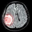
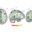
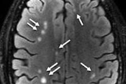
In other news, researchers from Walter Reed National Military Medical Center found that structural MRI could reveal additional information about brain scars in soldiers with mild traumatic brain injury (TBI) not found with techniques such as functional MRI (fMRI).
The researchers discovered T2-weighted hyperintense lesions in the brains of soldiers with mild TBI, a population that usually has normal MRI scans with fMRI sequences like perfusion MRI, diffusion-tensor imaging, and MR spectroscopy.
What do the abnormalities mean? The researchers aren't sure, but physicians at Walter Reed have found the results helpful in treating wounded warriors. Get the rest of the story by clicking here, or go to our MRI Community at mri.auntminnie.com.
PET/MRI for cardiac sarcoidosis
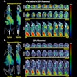
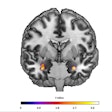
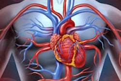
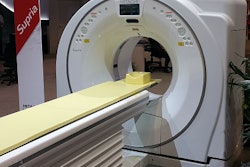
Finally, visit our Molecular Imaging Community for a new study on the use of PET/MRI for imaging cardiac sarcoidosis, an inflammatory heart condition that can lead to sudden cardiac death, but which is difficult to diagnose with modalities such as PET/CT. Read more by clicking here, or go to the community at molecular.auntminnie.com.


.fFmgij6Hin.png?auto=compress%2Cformat&fit=crop&h=100&q=70&w=100)


.fFmgij6Hin.png?auto=compress%2Cformat&fit=crop&h=167&q=70&w=250)
