Diffusion-tensor MRI (DTI-MRI) scans have shown significant brain changes in young football players after just one season of games and practice, even when they appear concussion-free, according to a new study published online October 24 in Radiology.
By using fractional anisotropy to track the movement of water molecules in the brain, researchers found microstructural alterations in the brain's white matter. Decreased fractional anisotropy values combined with cumulative exposure to impacts was associated with more random water movement, which has been associated with brain abnormalities in previous research.
"This study adds to the growing body of evidence that a season of play in a contact sport can result in brain changes at MR imaging, even in the absence of concussion," wrote lead author Naeim Bahrami, PhD, and colleagues (Radiology, October 24, 2016).
Repeated trauma
Repeated hits to the head in contact sports such as American football, soccer, and hockey have come under close scrutiny in recent years, as concerns escalate and evidence mounts about the long-term effects of concussion and related head trauma for athletes of all ages.
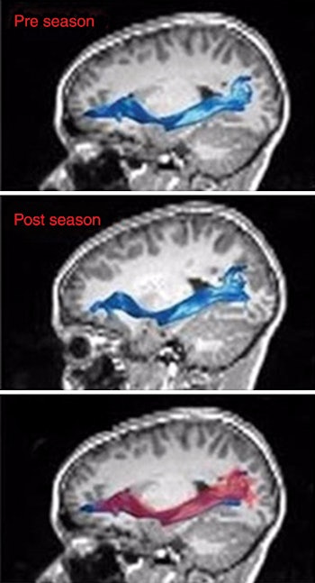
 MRI shows the left inferior fronto-occipital fasciculus before (top) and after (middle) the playing season. In an overlay of the two images (bottom), the red region illustrates the brain after the season, while the blue region represents the brain before the season. Image courtesy of Radiology.
A 2016 study by Churchill et al used MRI scans on college athletes with histories of concussion to show changes in brain size, blood flow, and structural white matter months and even years after injury. In a 2015 study by Albaugh et al, the researchers used MRI to show that concussions sustained by young ice hockey players were connected to subtle changes in the outer layer of the brain that controls higher-level reasoning and behavior.
This most recent paper comes from researchers at Wake Forest University, Virginia Tech, George Washington University, and University of Texas Southwestern. The collaborators targeted 25 male football players between the ages of 8 and 13 years (mean age, 11.72 years ± 1.05 years).
During the 2012 and 2013 season, head impacts were recorded using a Head Impact Telemetry System (HITS) and calculated as the combined probability risk-weighted cumulative exposure (RWECP) for a single season of play. All of the players' games and practices were also video recorded and reviewed to confirm the accuracy of the impacts.
Tracking the action
In the 2012 season, athletes' concussions were reported when a player, parent, and coach suspected the injury. In 2013, a certified athletic trainer was present for all games and practices and evaluated players suspected of having a concussion.
Study participants also underwent pre- and postseason evaluation with multimodal neuroimaging, including DTI-MRI of the brain on a 3-tesla MRI scanner (Skyra, Siemens Healthineers) with the aid of a high-resolution 32-channel head and neck coil (Siemens). The researchers collected 40 pairs of MR image sets from the 2012 and 2013 seasons that had the HITS data.
In particular, the group was interested in seasonal fractional anisotropy changes in three white-matter tracts -- the inferior fronto-occipital fasciculus, inferior longitudinal fasciculus, and superior longitudinal fasciculus.
"Studying specific fiber tracts within the brain, especially those of interest in mild traumatic brain injury, will assist in detecting the location of traumatic axonal injury as it relates to subconcussive head impact exposure," Bahrami and colleagues wrote.
Telltale signs
In 22 cases, there was a statistically significant relationship between RWECP results and decreased fractional anisotropy values in the left inferior fronto-occipital fasciculus (R2 = 0.4334, p = 0.003).
The statistically significant results were found in the whole left inferior fronto-occipital fasciculus (R2 = 0.433, p = 0.003), as well as the core (R2 = 0.3649, p = 0.007) and terminals (R2 = 0.5666, p = 0.001) of the left inferior fronto-occipital fasciculus.
"The statistically significant relationship between RWECP and change of fractional anisotropy in the left [inferior fronto-occipital fasciculus] suggests that an increase in subconcussive head impact exposure may have an effect on white-matter integrity in youth athletes, even in the absence of a clinically diagnosed concussion," the authors concluded.
There was the same statistically significant trend between RWECP and decreased fractional anisotropy values in the right superior longitudinal fasciculus (R2 = 0.2996, p = 0.042), but no such difference in the inferior longitudinal fasciculus.
Bahrami and colleagues believe that head impact exposure was the cause of decreased fractional anisotropy values of the whole and left inferior fronto-occipital fasciculus, as well as the right superior longitudinal fasciculus.
"The results of this study suggest that subconcussive impacts can result in changes in the white-matter microstructure of the inferior fronto-occipital fasciculus and the right inferior fronto-occipital fasciculus, as well as the right superior longitudinal fasciculus fiber bundles," they wrote. "This study adds to the growing body of evidence that a season of play in a contact sport can result in brain changes at MR imaging, even in the absence of concussion."
Cautionary note
The researchers were quick to add that none of the players showed any signs or symptoms of concussion from two seasons of games and practice.
"We do not know if there are important functional changes related to these findings, or if these effects will be associated with any negative long-term outcomes," corresponding author Dr. Christopher Whitlow from Wake Forest wrote in a prepared statement. "Football is a physical sport, and players may have many physical changes after a season of play that completely resolve. These changes in the brain may also simply resolve with little consequence. However, more research is needed to understand the meaning of these changes to the long-term health of our youngest athletes."


.fFmgij6Hin.png?auto=compress%2Cformat&fit=crop&h=100&q=70&w=100)


.fFmgij6Hin.png?auto=compress%2Cformat&fit=crop&h=167&q=70&w=250)


