An examination of MRI scans revealed that repeated head collisions incurred while playing football can deform the shape of white-matter tracts -- or bundles of nerve fibers -- in a child's brain, according to a presentation on Thursday at RSNA 2018.
"The years from age 9 to 12 are very important when it comes to brain development," said lead author Jeongchul Kim, PhD, from Wake Forest School of Medicine in Winston-Salem, NC. "The functional regions of the brain are starting to integrate with one another, and players exposed to repetitive brain injuries, even if the amount of impact is small, could be at risk."
Prior research has suggested that head impacts among youth football players can lead to functional changes in the brain, specifically in the brain's default mode network, which is critical for processing emotions.
In the study at hand, Kim and colleagues used a new MRI technique to visualize white-matter tracts in the brains of 48 boys, with the goal of seeking out any deformations in these nerve fiber bundles. Slightly more than half of the children had participated in a three-month youth football league.
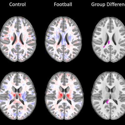
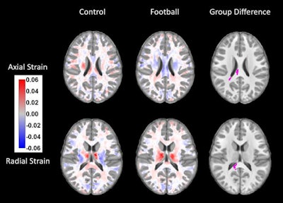
The MRI scans revealed that the brains of kids who played football had developed changes in the corpus callosum, the band of nerve fibers that integrates cognitive, motor, and sensory functions between the right and left hemispheres of the brain. The changes were marked by increased axial strain (fiber contraction) and radial strain (fiber expansion) in the brains of the football players, unlike in the brains of those who did not participate in contact sports.
 Brain MRI scans display statistically significant changes (violet) between the brains of kids before and after football season. Image courtesy of RSNA.
"The body of the corpus callosum is a unique structure that's somewhat like a bridge connecting the left and right hemispheres of brain," Kim said. "When it's subjected to external forces, some areas will contract, and others will expand, just like when a bridge is twisting in the wind. ... It's best to detect changes at the earliest possible time."
These findings are an early indication that repetitive blows to the head can result in alterations to the shape of the corpus callosum in youth football players.
Ultimately, the aim of this research is to provide guidelines for safe football play, Kim concluded. MRI may have a role in that process, specifically by helping physicians determine when a young athlete should be allowed to return to play after an injury to the head.


.fFmgij6Hin.png?auto=compress%2Cformat&fit=crop&h=100&q=70&w=100)


.fFmgij6Hin.png?auto=compress%2Cformat&fit=crop&h=167&q=70&w=250)


