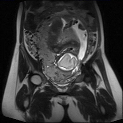
Further evidence of MRI's ability to confirm suspected placental anomalies or to assist in cases of equivocal ultrasound findings has come from Saudi Arabia. A study of pregnant women found that noncontrast MRI is a sensitive diagnostic test for detecting the increasingly common clinical problem of invasive placenta.
"Placenta accreta is a general term that means abnormal placentation, and it occurs when all or part of the placenta attaches abnormally to the myometrium," noted Dr. Metab Alkubeyyer, a consultant in body MRI and imaging informatics expert at King Khalid University Hospital and King Saud University Medical City in Riyadh. "It is associated with life-threatening bleeding during delivery. Incidence has been increasing steadily over the past years due to an increase in cesarean sections."
Detecting placenta invasion
Placenta accreta usually occurs in the lower anterior uterine segment, probably because of a deficiency of decidua at the level of the cesarean scar, he explained in an e-poster presented at ECR 2017 in Vienna. According to the depth of abnormal placentation, three grades are defined as the following:
- Placenta accrete (abnormal strong attachment to the myometrium)
- Placenta increta (abnormal invasion and penetration through the myometrium)
- Placenta percreta (invasion through the myometrium and uterine serosa)
Along with colleague Dr. N. Aldohayan, a third-year radiology resident at the same facility, he decided to assess the sensitivity and specificity of MRI to detect placenta invasion and to correlate the findings with intraoperative surgical findings.
They retrospectively reviewed 26 consecutive MRI studies of pregnant women (mean age 34.74 SD ± 5.5 years) admitted to King Khalid University Hospital between January 2012 and March 2016 with suspected invasive placenta. Multiplanar axial, coronal, and sagittal MR images were obtained with a slice thickness of 4 to 6 mm and a field-of-view of 30 to 40 cm using a 1.5-tesla machine (GE Healthcare) and single-shot fast-spin echo (SSFSE) and balanced steady-state free precession (B-SSFP) sequences.
Hospital staff obtained the images between the second and third trimesters, and coverage included the abdomen and pelvis. Patients with contraindications to MRI (pacemaker insertion, claustrophobia) were excluded from the study.
All images were reviewed by an expert radiologist blinded from intraoperative results. The researchers used Fisher's exact test for analysis between normal and invasive placenta groups with MRI outcome using web application GraphPad QuickCalcs (graphpad.com).
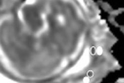
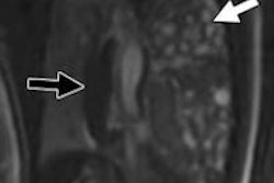
The MRI signs and criteria for invasive placenta included intraplacental T2 dark bands, lobulation, and heteogeneity of placenta, and direct invasion to adjacent structures.
In their analysis, the researchers did not include five of the 26 patients for whom intraoperative notes were missing. A total of 17 of the remaining 21 patients (81%) had a cesarean section, and 20 (95%) of the cases had placenta previa. The intraoperative results showed four (19%) patients with invasive placenta, compared with 17 (81%) who had normal placenta.
Placenta invasion in MRI was positive in eight (38%) patients and negative in 13 (62%) cases. MRI showed 100% sensitivity and 76.47% specificity in the detection of invasive placenta with significant statistical association (Fisher's exact test p = 0.012) between patients with invasive placenta and positive MRI outcome.
The routine method used to evaluate the placenta remains screening ultrasound at 18 to 20 weeks of pregnancy, but it may be inconclusive or equivocal in cases of a difficult visualization of the placenta due to the patient's body habitus or a posterior placental implantation. The main challenge with MRI, however, is that the signs may be subtle and difficult to detect, especially if the radiologist has limited experience. Determining the diagnostic value of specific MR features for detecting suspected placental invasion can differ according to the radiologist's experience.
Overall, MRI can achieve an acceptable level of specificity when the appropriate technique is used, Alkubeyyer and Aldohayan stated.
To view the authors' full ECR 2017 e-poster on the European Society of Radiology's website, click here.
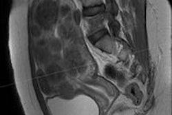
Editor's note: MR image on home page shows major placenta previa. Image courtesy of Dr. Jamal Alkoteesh, Al Ain Hospital in United Arab Emirates.


