Researchers have developed a way to enhance MR images to aid large-scale studies designed to show how strokes affect different people and how patients respond to treatment.
Scans performed on stroke patients at hospitals often have too low of a resolution for research analyses using various algorithms. Researchers usually obtain images with much higher resolution for use in scientific studies.
The group from the Massachusetts Institute of Technology wanted to investigate a way to utilize the large amount of these lower-quality scans. The new approach involves filling in missing data from each patient scan by taking information from the entire set of MRI scans and using it to recreate omitted anatomical features.
"The key idea is to generate an image that is anatomically plausible, and to an algorithm looks like one of those research scans, and is completely consistent with clinical images that were acquired," said senior author Polina Golland, PhD, a professor of electrical engineering and computer science, in a statement. "Once you have that, you can apply every state-of-the-art algorithm that was developed for the beautiful research images and run the same analysis, and get the results as if these were the research images."
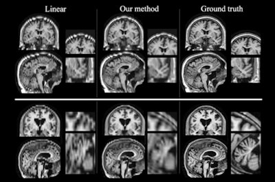
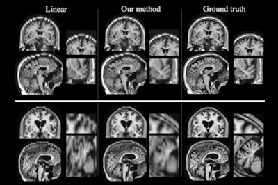 MR images produced by a newly developed method (center column) more closely resemble high-resolution scans (right column). Images courtesy of Golland et al.
Golland is scheduled to present the study the week of June 25 at the Information Processing in Medical Imaging conference in Boone, NC.
Using these scans, researchers could study how genetic factors influence stroke survival or how people respond to different treatments. They could also use this approach to study other disorders such as Alzheimer's disease.


