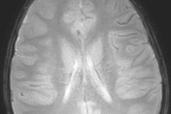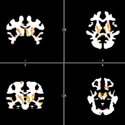
An MRI technique aimed at quantifying changes in myelin content revealed that athletes who have a mild traumatic brain injury (TBI) can experience changes to their brain white matter that are still evident months later, according to a preliminary study published online May 1 in the Journal of Neurosurgery.
Seeking to develop better diagnostic and prognostic tools for mild TBI, a multi-institutional team of researchers used an emerging multicomponent relaxation MRI technique -- multicomponent driven equilibrium single-pulse observation of T1 and T2 (mcDESPOT) -- to scan more than 20 college athletes. The subjects included football and rugby players with a recent mild TBI, as well as healthy athletes from noncontact sports.
The researchers found that myelin water fraction -- used as a surrogate MRI measure of myelin content -- was higher in the brains of the athletes with TBI than in the normal controls. Changes in the myelin water fraction represent changes in the amount of myelin, which acts as a sheath covering the axons of neurons and facilitates the transmission of electrical impulses throughout the brain, according to the group. The higher the myelin water fraction, the more myelin is present.
"We were surprised by the finding of increased myelin in the contact sports players compared with noncontact sports players at baseline and three months after injury," said lead author Dr. Heather Spader from Joe DiMaggio Children's Hospital in Hollywood, FL, in a statement from the journal. "Using the mcDESPOT sequence, we can see that there is a remyelination process after an injury."
The study included 23 male college athletes from Brown University; 13 were rugby and football players who had a recent mild TBI, while the remaining 10 were healthy athletes from noncontact sports who served as age-matched controls. The contact sport players underwent MRI -- including mcDESPOT and additional sequences -- within 72 hours of the injury and again three months later. The control subjects received a single baseline imaging session with the same protocol.
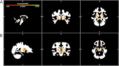 Myelin water fraction maps comparing the brains of contact sport players three months after mild TBI with the noncontact sport player baseline. A: Sagittal (left), coronal (center), and axial (right) maps at the level of the thalamus showing increased myelin water fraction (p < 0.05, familywise error corrected) in the anterior and posterior corpora callosa, thalamus, and midbrain. B: Sagittal (left), coronal (center), and axial (right) maps showing significant myelin water fraction increases (p < 0.05, familywise error corrected) in the midbrain and temporal lobes (left > right temporal lobe). Images courtesy of the American Association of Neurological Surgeons.
Compared with the baseline images from the healthy athletes, the contact sport athletes showed significantly higher myelin water fractions at baseline in the bilateral basal ganglia, anterior and posterior corpora callosa, left corticospinal tract, and left anterior and superior temporal lobe. What's more, they also demonstrated significantly higher myelin water fractions three months later in the bilateral basal ganglia, anterior and posterior corpora callosa, and left anterior temporal lobe, as well as in the bilateral corticospinal tracts, parahippocampal gyrus, and bilateral juxtaposition areas, according to the researchers.
The contact sport athletes also had higher overall myelin water fraction at the time of injury and three months later, compared with the healthy athletes. The increases in overall myelin water fraction were greatest in the bilateral basal ganglia and deep white matter, while decreases were seen in more superficial white matter, the researchers found.
Spader and colleagues noted, though, that increased myelin alone is not necessarily a good thing. Animal studies have shown that remyelination following mild TBI may result in disorganized and less functional myelin. Ominously, a related investigation by the researchers found that the sites of increased myelin water fraction in contact sport players corresponded on PET imaging to sites of brain changes in patients suspected of having chronic traumatic encephalopathy (CTE).
Nonetheless, the researchers said it's too soon to draw definitive conclusions from this preliminary research. They recommended additional studies to determine a true baseline for myelin water fraction in athletes before an injury occurs, rather than using the surrogate baseline myelin water fraction from players of noncontact sports.
"The next question is to determine if the increased myelin leads to the formation of a type of scar tissue that can cause disorganized signaling in the brain and which can eventually lead to an increased susceptibility to neurodegenerative disorders such as dementia," Spader said.


