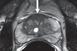
Multiparametric MRI is a valuable option for monitoring patients who have undergone MRI-guided focal laser ablation for prostate cancer, according to a study published online July 11 in the American Journal of Roentgenology.
Led by Dr. Charles Westin from the department of radiology at the University of Chicago, the researchers evaluated 27 men with clinical category T1c-T2a prostate cancer, a prostate-specific antigen (PSA) level of 10 mg/mL or less, and a Gleason score of 7 or less. The subjects also had undergone prostate biopsy before and after focal laser ablation to treat a total of 36 lesions.
In addition, 3-tesla MRI scans (Achieva, Philips Healthcare) were performed before ablation, immediately after the procedure, and again three and 12 months after ablation to monitor treatment. The multiparametric MRI protocol included T2-weighted imaging and diffusion-weighted imaging (DWI). The mean lesion size before ablation was 6.3 mm (range: 3-13 mm).
Three months after ablation, MRI-guided biopsy of ablation zones revealed that 26 men (96%) had no evidence of cancer. Twelve months after ablation, however, biopsy showed that 10 men had recurrent cancer; three of those patients had cancer at the ablation site.
"Typically, in the absence of residual cancer, the ablation zone was not seen or was seen as a nonenhancing focus on imaging examinations performed 12 months after ablation," the authors noted. "In conclusion, multiparametric MRI can show postablation changes in the prostate and can be a valuable tool for the follow-up of patients who have undergone MRI-guided focal laser ablation."


