Wednesday, November 28 | 3:30 p.m.-3:40 p.m. | SSM13-04 | Room E353C
Patients with cancer may better understand their disease and treatment options by examining personalized 3D models made with 3D printing or augmented reality (AR) technology, according to this Wednesday presentation.Clinicians often explain to patients the nature of their disease and the surgical plan to manage it using conventional 2D imaging as a reference. Researchers from NYU Langone Medical Center have turned instead to advanced visualization technologies to further enhance patient education.
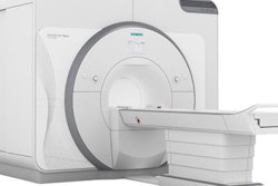
To that end, the group led by presenter Nicole Wake, PhD, acquired the MRI scans of 73 patients with renal cancer or prostate cancer who were scheduled to undergo surgical treatment. Wake and colleagues created patient-specific 3D virtual models using AR technology, as well as 3D-printed kidney and prostate models based on these data. Next, they allowed the patients to view their MRI scans or one of the 3D models before completing questionnaires.
Reviewing the patients' responses, the researchers found that the more enhanced and intuitive visualization afforded by the 3D-printed and AR models improved patient understanding substantially. The patients demonstrated statistically significant improvements in their knowledge of the disease, cancer size, cancer location, and treatment plan when studying the 3D-printed models. Looking at AR models also led to improvements in the patients' awareness of the size of the cancer.
"Being able to see and hold these tangible 3D models brings everything together for patients, allowing them to immediately understand their disease," Wake told AuntMinnie.com. "In addition, using these models to explain the surgical procedure ultimately helps patients make improved surgical decisions."


