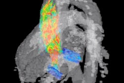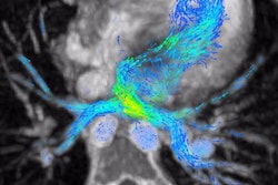
An automated heart valve-tracking algorithm halved the amount of time and variability involved in blood-flow quantification with 4D MRI, according to an article published online October 30 in Radiology. The improved efficiency may help convince clinicians that cardiac 4D MRI is useful for assessing valvular heart disease.
The diagnosis and management of valvular heart disease relies on the accurate quantification of blood-flow volumes through heart valves. Blood-flow quantification is most commonly achieved by analyzing standard MRI scans or echocardiography images, but recent studies have shown that using 4D MRI data leads to more accurate measurements, noted first author Dr. Vivian Kamphuis and colleagues from Leiden University Medical Center in the Netherlands.
Despite the superior accuracy of 4D MRI over conventional MRI, clinicians seldom perform the advanced imaging technique, as it requires a time-consuming and often highly variable manual task known as heart valve tracking, the authors wrote.
Seeking to overcome this limitation, Kamphuis and colleagues developed a valve-tracking algorithm compatible with 4D MRI software (Caas MR 4D Flow, Pie Medical Imaging) that was capable of automatically identifying heart valves and tracking their blood flow during 4D MRI.
The researchers acquired the data of 114 patients with heart valve disease who underwent 4D MRI at their institution, both with and without their automated valve-tracking algorithm. They found that automated valve tracking not only produced blood-flow measurements considerably faster than manual tracking but also led to more consistent results.
| Manual vs. automated valve tracking for 4D MRI blood-flow quantification | ||
| Manual valve tracking | Automated valve tracking | |
| Time for blood-flow analysis | 25 minutes | 14 minutes |
| Variability in blood-flow measurements | 9.8% | 4.9% |
What's more, automated valve tracking would have led to the reclassification of disease severity for as many as 28 patients if clinicians had used the technique in the initial evaluation. This suggests its potentially significant role in clinical decision-making for cases of heart valve disease, the authors wrote.
"Cardiac [4D] MRI offers several advantages for quantifying the severity of tricuspid and mitral regurgitation," wrote Dr. Christopher François from the University of Wisconsin School of Medicine and Public Health in an accompanying editorial. "By using automatic valve-tracking methods, the adoption of cardiac [4D] MRI in the evaluation of patients with tricuspid and mitral regurgitation is expected to increase."
Applying machine learning to 4D MRI may improve the accuracy of such automated valve-tracking algorithms even more -- further reducing analysis times and variability, he concluded.


