Planning a complex surgery for patients with congenital heart disease may require multiple advanced visualization techniques, say researchers from California, who used both 4D flow MRI and 3D printing for a case study recently published in the World Journal for Pediatric and Congenital Heart Surgery.
In the past several years, 4D flow MRI has become increasingly popular for imaging patients who require a Fontan procedure, a surgery for pediatric patients who have only a single functional heart ventricle, noted first author Dr. Thomas Carberry from the University of California, San Diego and colleagues. The technology allows for the quantification of essential clinical measures -- such as pulmonary blood-flow distribution -- that can help clinicians make appropriate management decisions.
Various groups have also recently taken to using 3D printing to plan and simulate surgeries for congenital heart diseases.
Taking advantage of these emerging technologies, Carberry and colleagues used both 4D flow MRI and 3D printing to plan revision surgery for a 10-year-old girl whose oxygen levels remained low despite having undergone a series of heart surgeries that included a Fontan procedure (World J Pediatr Congenit Heart Surg, January 10, 2019).
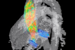
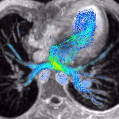
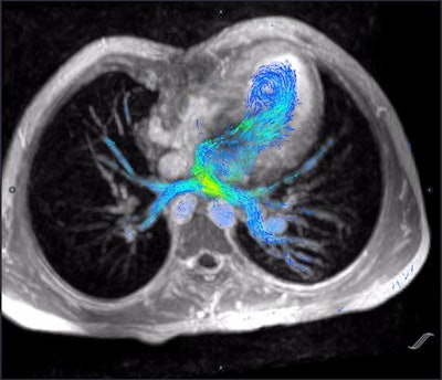 Cardiac 4D flow MRI scan showing prepulmonary vein blood flow. Image courtesy of Dr. Albert Hsiao, PhD.
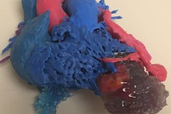
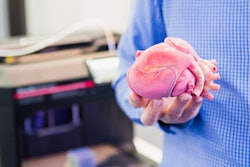
The researchers began by acquiring cardiac 4D flow MRI and CT scans of the patient. They used the CT scans to create one 3D-printed heart model with a standard 3D printer (Fortus 400, Stratasys) and a second model using a type of material that replicated the elastic texture of cardiac tissue. They used the 4D flow MRI scans to quantify blood flow in the heart vasculature.
The 3D-printed heart models and 4D flow MRI data allowed the clinicians to assess blood flow as well as examine the patient's heart anatomy in great detail and from several different perspectives. This helped them to better understand the anatomic relationships between the heart and surrounding structures during preoperative planning, and the models also proved beneficial in explaining surgical options to the patient's family.
After examining the imaging data and models, a surgical team was able to determine the most effective way to approach the heart revision surgery. Ultimately, the surgery significantly improved the patient's oxygen saturation levels, with healthy blood flow distribution confirmed on postoperative 4D flow MRI scans.
"Three-dimensional printing is emerging as a useful technology in congenital heart disease, and this case shows an example of its use in conjunction with 4D flow MRI for complex cardiac surgery," the authors wrote.
"Ideally, as 3D printing becomes more cost-effective and available, this can be used more readily in conjunction with other novel presurgical imaging technologies," they concluded.


