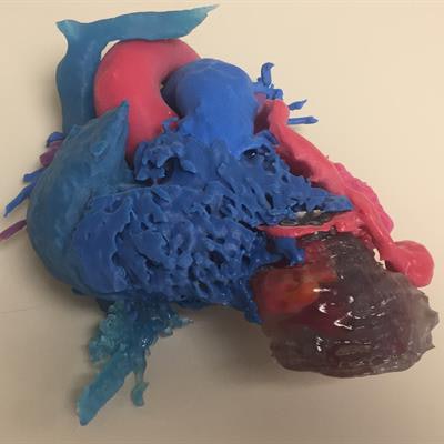
A 3D-printed heart model based on patient MR images helped physicians identify two uncommon cardiac anomalies in a case report published online November 11 in Clinical Imaging. The technique may be a viable alternative to more invasive procedures, such as cardiac catheterization, researchers say.
A group of physicians from Washington University School of Medicine in St. Louis led by Dr. Mahati Mokkarala was in charge of caring for a 25-year-old pregnant woman who presented with palpitations and near syncope. Standard MRI revealed the presence of multiple anomalies in the patient's heart but with limited visualization of the underlying anatomy.
The researchers suspected that the patient's symptoms were likely a result of two distinct, rare pathologies based on the MR images: a double-chambered right ventricle or a coronary cameral fistula. Double-chambered right ventricle is a congenital heart anomaly that can result in significant ventricular obstruction and typically requires myomectomy, whereas coronary cameral fistulas often require transcatheter closure or a different surgical intervention.
Due to the complexity of the patient's pathology, Mokkarala and colleagues decided to create a 3D-printed heart to improve visualization of the patient's heart following the completion of pregnancy and delivery. To that end, they acquired gadolinium-enhanced MR images of the patients using a whole-body 1.5-tesla scanner with a high-resolution gradient-recalled echo sequence.
Next, they segmented the heart and surrounding anatomy from the MR images, generated virtual 3D models using the segmentations, and then 3D printed a patient-specific, color-coded heart model.
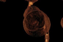
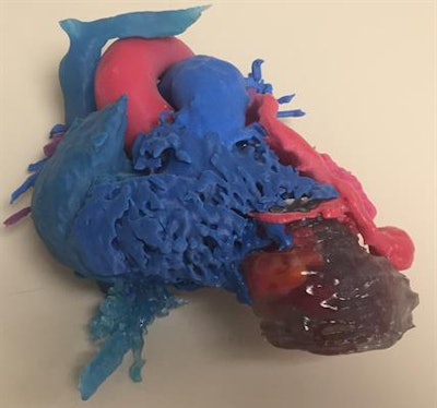 3D-printed heart based on the MR images of a woman with a coronary cameral fistula connected to a double-chambered right ventricle. Image courtesy of Dr. David Ballard.
The researchers found that the 3D-printed heart clearly showed the anatomic relationship between the double-chambered right ventricle and the coronary cameral fistula, which were found to be connected. The model also showed an enlarged left coronary artery and small channels through the fibrous band connecting the right heart chambers. Postpartum cardiac catheterization subsequently confirmed these findings.
"For this patient, 3D computer modeling and printed structures were useful for visualizing and contextualizing complicated anatomy including the connection between the coronary cameral fistula and double-chamber right ventricle," the team wrote.
Clinicians generally have to rely on multiple modalities -- including echocardiography, MRI, and cardiac catheterization -- to diagnose and plan treatment for the two cardiac abnormalities. 3D printing technology may allow for diagnosis and treatment planning of these conditions and other right ventricle pathology without the need to use as many modalities, they noted.
"This case highlights the utility of cardiac MRI and 3D printing technology in the diagnosis of a patient with unusual right ventricle and coronary anatomy," the group concluded.


