Cardiac 4D flow MRI can quantify blood flow in the ascending aorta (AAo) and main pulmonary artery (MPA) during strenuous exercise, offering potential as a useful modality for evaluating right-sided heart dysfunction, according to research published online June 18 in Radiology: Cardiothoracic Imaging.
In a pilot study, a team of researchers led by Jacob Macdonald, PhD, of the University of Wisconsin prospectively evaluated nine patients with 4D flow MRI at rest and during strenuous exercise. They found that the technique was highly repeatable and showed excellent correlation between observers for measuring flow in the AAo and MPA.
On the downside, quantification of kinetic energy (KE) in the left and right ventricle had poor interobserver correlation.
"With further development, biventricular 4D flow exercise cardiac MRI can fulfill a valuable clinical role in evaluating patients with right-sided heart dysfunction, a group not well-addressed by stress echocardiography," the authors wrote.
Using a Discovery 750 3-tesla MR scanner (GE Healthcare) with an eight-channel cardiac coil, the researchers prospectively performed 4D flow MRI on nine healthy volunteers at rest and during exercise with an MRI-compatible exercise stepper between March 2016 and July 2017. 4D flow MRI was performed with a radially undersampled 4D flow sequence, and the acquisition was retrospectively gated for both respiratory and cardiac motion.
The Mimics software (version 17, Materialise) was used to segment the AAo and MPA on the time-averaged, complex difference 4D flow MR images. Flow was then quantified using the Ensight software (version 10, Ansys) and MATLAB (MathWorks).
The researchers assessed interobserver variability by comparing measurements from the original observer, who had five years of 4D flow postprocessing experience, with that of a second observer with two years of 4D flow postprocessing experience.
Although signal-to-noise ratio decreased by an average of 16% for exercise exams, the researchers concluded that there was no significant difference in image quality between the rest and exercise exams for the vessel segments that were investigated. Internal flow consistency analysis showed average relative differences of 10% at rest and 16% during exercise. With intraclass correlation coefficients (ICCs) of more than 0.9, both AAo and MPA measurements also demonstrated excellent interobserver and intraobserver reliability.
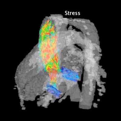
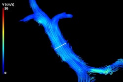
Representative pathline animations derived from 4D flow images in the right ventricle during rest (left) and exercise (right). Noticeably higher velocities are present in the right ventricle and main pulmonary artery during exercise. Video courtesy of Radiology: Cardiothoracic Imaging.
However, kinetic energy measurements showed significant increases -- approximately 30% -- in average relative differences, as well as poor (ICCs < 0.5) intraobserver and interobserver correlation.
"Four-dimensional flow MRI can quantify increases in flow in the AAo and MPA during strenuous exercise and is highly repeatable," the authors wrote. "KE had reduced repeatability because of suboptimal segmentation methods and requires further development before clinical implementation."
The study results are promising, said Michael Markl, PhD, and Jeesoo Lee, PhD, of Northwestern University in an accompanying commentary.
"A combination of cardiopulmonary exercise testing with 4D flow MRI may enable new physiologic insights into the complex volumetric changes in in vivo 3D blood flow dynamics in healthy participants and patients with cardiovascular disease," they wrote. "This study is an important first step to demonstrate the feasibility of this approach. Future studies in larger cohorts of patients are needed to confirm these promising findings and further explore applications of 4D flow during rest and exercise to assess the impact of demographic factors (age, sex), as well as disease, on cardiovascular hemodynamics."


