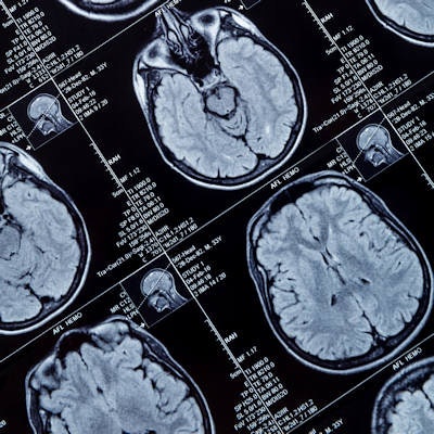
Results of a type of MRI protocol called neuromelanin-sensitive MRI (NM-MRI) could help diagnose psychosis and provide an indicator of psychosis severity, according to a study published February 18 in the Proceedings of the National Academy of Sciences.
The approach uses the MRI signal to target neuromelanin, a pigment that is created within dopamine neurons of the brain's substantia nigra. Dopamine plays a key role in mental and neurological disorders, such as schizophrenia and Parkinson's disease; therefore, finding a method to measure dopamine activity in the brain could help to advance diagnoses and subsequent treatment.
"The main advantages of this technique are that, compared to other established and more direct measures of dopamine function, neuromelanin-sensitive MRI does not involve radiation or invasive procedures," said study co-author Dr. Guillermo Horga, PhD, an assistant professor of psychiatry at Columbia University in New York. "This advantage makes it more suitable for pediatric populations and for repeated scanning, which could be useful to monitor the progression of illness or response to treatment."
NM-MRI also can be performed on most clinical scanners and requires only a short scan time. Another benefit is the high anatomical resolution, which is considerably better than PET and allows for better assessment of the substantia nigra's functions or dysfunctions.
The researchers tested the suitability of NM-MRI for this use by seeing if it could accurately detect regional variations in neuromelanin concentration in subjects with no evidence of neurodegeneration of the substantia nigra. They compared NM-MRI neuromelanin measurements to chemical measurements of neuromelanin in postmortem brain tissue and observed a higher NM-MRI signal across all tissue. The greater signal was associated with higher concentrations of neuromelanin, which confirmed NM-MRI's efficacy in this task.
They then used NM-MRI data from patients with and without Parkinson's disease to determine variations in neuromelanin in smaller anatomical subregions of the substantia nigra. Horga and colleagues discovered decreases in NM-MRI signal in the lateral, posterior, and ventral areas of the substantia nigra among patients with Parkinson's. The results again confirmed that NM-MRI can discern neuromelanin variability within this brain region.
Finally, the researchers examined the link between NM-MRI signal and psychosis severity. They found an association between severe symptoms of psychosis and higher NM-MRI signals in the nigrostriatal pathway of individuals with schizophrenia and those at risk for schizophrenia. Again, NM-MRI revealed the dopamine system dysfunction, which would support the technique's results as a potential biomarker for psychosis.
"We are now extending this work to see if we can detect abnormalities in neuromelanin signal that help us predict which individuals are more likely to develop a psychotic disorder among those who already show early symptoms of psychosis," Horga added. "We are also interested in exploring whether neuromelanin-sensitive MRI could be used in the future to determine who might best benefit from dopaminergic treatments."


