A combination of 7-tesla MRI and 3D printing technology has revealed that patients with a growing number of chronic active brain lesions are also more likely to have more debilitating forms of multiple sclerosis (MS), including individuals receiving standard treatment. The findings were published online August 12 in JAMA Neurology.
The ultrahigh field strength of 7-tesla MRI has proved effective in identifying otherwise difficult-to-find lesions in the brains of patients with MS -- leading to new insight into the relatively elusive disease. Of note, recent research from the U.S. National Institutes of Health (NIH) linked the presence of dark-rimmed spots, or chronic active lesions, on 7-tesla MRI scans with various disabilities in individuals affected by MS.
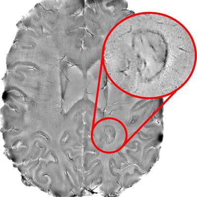
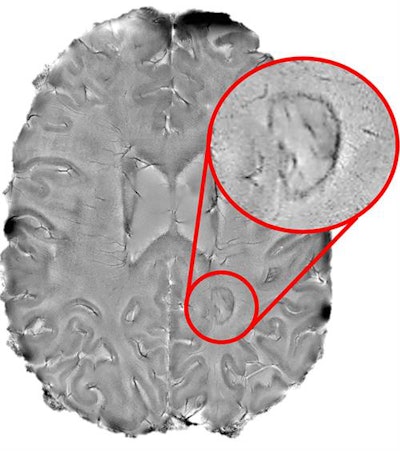 Chronic active lesions that appear as dark-rimmed spots (red circle) on brain MRI scans indicate "smoldering" inflammation, a sign of debilitating multiple sclerosis. Image courtesy of the NINDS Reich lab.
"Figuring out how to spot chronic active lesions was a big step and we could not have done it without the high-powered MRI scanner provided by the NIH," first author Dr. Martina Absinta, PhD, from the National Institute of Neurological Disorders and Stroke (NINDS) said in a statement from the NIH. "It allowed us to then explore how MS lesions evolved and whether they played a role in progressive MS."
Expanding upon this earlier research, Absinta and colleagues investigated the long-term relationship between chronic rim lesions detected with 7-tesla or 3-tesla MR imaging and MS-related disabilities in 192 MS patients. Approximately 34% of the patients had one to three chronic rim lesions and 22% had at least four rimmed lesions, despite receiving conventional drug therapy.
Overall, the likelihood of clinically progressive MS was 1.6-fold higher in patients with four or more chronic rim lesions, compared with those who did not have a rimmed lesion (p = 0.03).
In addition, patients who had four or more rimmed lesions were affected by motor and cognitive disabilities at a much younger age, according to the researchers. This pattern was especially pronounced in patients younger than 50 years old: The prevalence of severe MS was 3.2 times greater in patients with four or more rimmed lesions than in those without rimmed lesions in this younger subgroup.
Advanced MR imaging enabled the detection of chronic active lesions and thereby helped determine which patients with MS were most susceptible to aggressive forms of the disease, said senior investigator Dr. Daniel Reich, PhD, director of the NINDS translational neuroradiology division.
Following this prospective study, Absinta and colleagues acquired postmortem brain MRI scans of a 50-year-old man with progressive MS. They converted the scans into virtual 3D models using computer software. Next, they used these models to create a digital template of a brain slicer, or cutting box, which they printed out using a 3D printer.
The resulting 3D-printed cutting box allowed the researchers to slice the brain into ideal sizes for histological examination. Their analysis of the pathology slides confirmed that the presence of multiple rimmed lesions was associated with considerable decreases in white-matter volume and basal ganglia size in the patients' brains.
The researchers conducted yet another study assessing long-term changes to brain lesions in 23 MS patients who underwent 10 years of annual MRI exams. They found that the chronic rim lesions expanded at an annual rate of 2.5% (including in patients undergoing anti-inflammatory drug therapy), whereas lesions without rims shrank by 4.7% per year.
"Our results support the idea that chronic active lesions are very damaging to the brain," Reich said. "We need to attack these lesions as early as possible. The fact that these lesions are present in patients who are receiving anti-inflammatory drugs that quiet the body's immune system also suggests that the field of MS research may want to focus on new treatments that target the brain's unique immune system."


