MR images indicate that methylphenidate, also known as Ritalin, can affect certain areas of white matter in the brains of preteen children who have attention deficit hyperactivity disorder (ADHD), according to a Dutch study published online August 13 in Radiology.
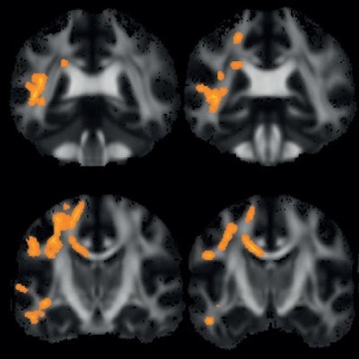
The researchers used diffusion-tensor MR imaging (DTI-MRI) to observe greater fractional anisotropy values in children who were treated with the drug, but they found no such reactions in adults with ADHD who also took the medication or in groups of similarly matched children and adults who took placebos.
The findings are among the first steps in trying to determine the degree to which treatment with the drug to control ADHD could affect the maturation of young brains.
"A strength of our current study was its prospective design, in which the effects of confounders, such as age and sex, are likely to be relatively small," wrote the study authors, led by Dr. Cheima Bouziane, from Amsterdam University Medical Centers. "The selective inclusion of stimulant treatment-naive study participants also was critical for addressing our objective."
Methylphenidate is marketed by Novartis under the brand name Ritalin. The drug is commonly prescribed for ADHD, which is associated with abnormal development of white-matter tracts in the brain. Previous studies have shown the drug to be effective in 60% to 80% of ADHD cases, but the research is retrospective and "possible confounding effects of medication were not taken into account," Bouziane and colleagues noted.
One preclinical MRI study found increased fractional anisotropy in the corpus callosum of methylphenidate-treated adolescent rats but no changes in adult rats given a saline solution. The result is noteworthy because higher fractional anisotropy values are an indication of disrupted water flow in the brain's white matter. Therefore, the drug's efficacy could depend on the recipient's age, with the question of how the greater fractional anisotropy levels might affect brain development in young ADHD patients.
In the current study, the researchers from the Netherlands hypothesized the same preclinical results could apply to humans, so they devised a 16-week prospective, randomized, controlled trial with 25 boys who received methylphenidate and 25 boys who were given a placebo (age range, 10-12 years). In addition, 24 men received the drug, and 24 men were in a placebo group (age range, 23-40 years). All DTI-MRI studies were performed on a 3-tesla scanner (Philips Healthcare) with an eight-channel head coil and body coil at baseline and one week after the end of treatment to ensure drug "washout."
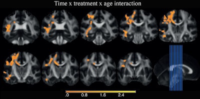
At the 16-week follow-up mark, a comparison of baseline and post-treatment diffusion-tensor images revealed significantly greater fractional anisotropy values among the boys treated with the drug. By comparison, the researchers found no significant changes in fractional anisotropy among ADHD adults given the drug or in either of the two placebo groups.
 Voxelwise fractional anisotropy analysis shows significant changes relative to age and MHP treatment. Coronal sections (left) correspond to the sagittal section locations in the image in the bottom row far right. The areas in which the difference between baseline and after treatment in children treated with methylphenidate are greater than adults treated with the drug are color coded orange. They are located in parts of the left superior longitudinal fasciculus, inferior longitudinal fasciculus, inferior fronto-occipital fasciculus, and the corpus callosum truncus. Images courtesy of Radiology.
The Dutch researchers recommended additional studies to investigate whether their "findings can be extrapolated to the female sex and to young/older children and/or adolescents, with more advanced protocols ... which would allow researchers to distinguish additional white-matter details, such as fiber crossings, which cannot be reliably estimated otherwise."


