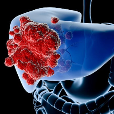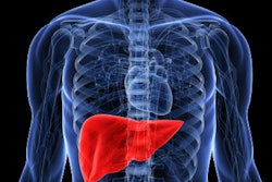
Tomoelastography -- an MRI technique that enables visualization of the mechanical properties of tumors -- can accurately distinguish between benign and malignant liver lesions, according to research from Germany published online September 24 in Cancer Research.
A team of researchers led by Dr. Ingolf Sack of Charité University Medicine Berlin used tomoelastography to show that hepatic malignancies contain tissues with both stiff and fluid properties, while surrounding tissues are predominantly solid.
"Our findings regarding these unusual mechanical properties may be indicative of a general pattern for cancer growth," said lead author Mehrgan Shahryari in a statement. "Our findings may help researchers to develop entirely noninvasive means of distinguishing between benign and malignant lesions."
With tomoelastography, patients are exposed to acoustic waves for approximately five minutes during an MR scan. Next, the spread of mechanical waves within the liver is mapped, revealing changes in the solid-fluid tissue properties of soft tissues, according to the researchers.


.fFmgij6Hin.png?auto=compress%2Cformat&fit=crop&h=100&q=70&w=100)


.fFmgij6Hin.png?auto=compress%2Cformat&fit=crop&h=167&q=70&w=250)


