Functional MRI (fMRI) has shown that prenatal exposure to opioids may alter brain function in babies, according to research scheduled to be presented at the upcoming RSNA 2019 meeting in Chicago.
A team of researchers led by Dr. Rupa Radhakrishnan of Indiana University in Indianapolis used resting-state fMRI to study 16 full-term infants. They found that infants with prenatal opioid exposure had significant differences in the way the amygdala connected to various brain regions.
Prenatal exposure to opioids may result in the baby having a group of conditions known as neonatal abstinence syndrome (NAS), and in utero exposure is believed to have lasting consequences on brain development and behavior, according to the researchers.
What's more, the syndrome requires prolonged hospital stays, monitoring, and, in severe cases, additional treatment with opioids. Understanding how opioids affect the developing brain would be an important step in the early identification and management of NAS and improving neurodevelopmental and behavioral outcomes in these children, the researchers noted.
"Little is known about brain changes and their relationship to long-term neurological outcomes in infants who are exposed to opioids in utero," Radhakrishnan said in a statement from the RSNA. "Many studies have looked at the impact of long-term opioid use on the adult and adolescent brain, but it is not clear whether social and environmental factors may have influenced those outcomes. By studying infants' brain activity soon after birth, we are in a better position to understand the effect of opioids on the developing brain and explain how this exposure could influence long-term outcomes in the context of other social and environmental factors."
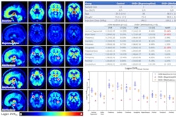
The group of researchers, which included obstetricians, neonatologists, psychologists, and imaging scientists, studied 16 full-term infants -- eight with prenatal opioid exposure and eight without. Anatomical and functional MRI scans were performed on the babies while they were sleeping.
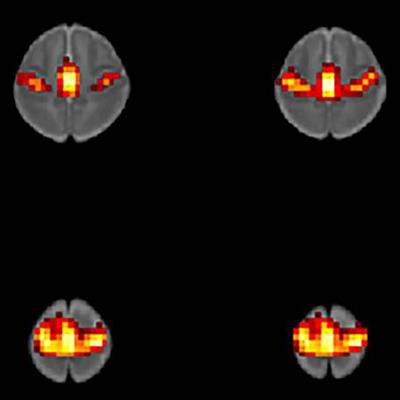
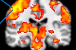
After creating brain maps and applying regions of interest for the left and right amygdala, the researchers found significant differences in the way the amygdala connects to different brain regions.
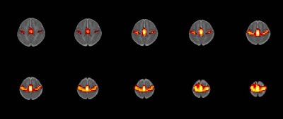
 fMRI scan of resting-state networks acquired as part of the study. Image courtesy of the RSNA.
Larger and long-term outcome studies are now underway to better understand the functional brain changes in prenatal opioid exposure and their associated long-term development outcomes, according to Radhakrishnan.
"Although our early results showed differences between the two groups in a small study sample, it is very important that we further investigate and validate these findings in larger studies," Radhakrishnan concluded. "To identify the best methods for managing NAS and improving long-term outcomes in these infants, it is critical to understand changes in brain function that may result from exposure to opioids prenatally."


