Air pollution is not only bad for the lungs, it could be bad for the brain as well -- especially in children. Brain MR images showed reduced gray-matter volume in kids who were exposed to high levels of traffic-related air pollution since birth, according to a January 24 study in PLOS One.
The findings are particularly noteworthy because the brain regions with the greatest reductions -- the frontal and parietal lobes and the cerebellum -- deal with cognitive performance, sensory perception, sight, and hearing and could slow a child's natural development.
"The results of this study, though exploratory, suggest that where you live and the air you breathe can affect how your brain develops," said lead study author Travis Beckwith, PhD, a research fellow at Cincinnati Children's Hospital Medical Center, in a statement. "While the percentage of loss is far less than what might be seen in a degenerative disease state, this loss may be enough to influence the development of various physical and mental processes."
The subjects came from the prospective Cincinnati Childhood Allergy and Air Pollution Study, which recruited children younger than age 6 months to examine their early exposure to traffic-related air pollution and followed them to age 12 to see if they developed health issues such as allergies, asthma, and neurological dysfunction. Eligibility criteria included a residence less than a quarter-mile or more than 1 mile from a major highway and at least one biological parent with a tendency to develop allergies.
The children were clinically examined at 1, 2, 3, 4, 7, and 12 years old. Parents completed questionnaires about the children's health, housing situation, and residential history. When the children were age 12, researchers conducted "direct and indirect assessments of intelligence, reading ability, executive function, mental health, and other neurodevelopmental outcomes," the authors noted.
Exposure to traffic-related air pollution was estimated by taking data from 27 sites in the greater Cincinnati area. Air-quality information was inserted into a model to determine carbon levels from diesel emissions and measure elemental carbon attributable to traffic (ECAT). Based on the level of carbon concentration, children were deemed to have high or low exposure to air pollution. The mean estimated ECAT exposure at birth was twice as great (0.56 ± 0.13 µg/m3) in the highly exposed group as the lower-exposed group (0.27 ± 0.02 µg/m3).
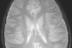
Over the course of the study, a total of 135 children completed 3-tesla MRI scans (Achieva, Philips Healthcare) with a 32-channel head coil. The researchers then used voxel-based morphometry to examine differences in regional brain volume and cortical thickness between the two groups.
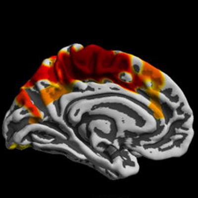
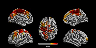
The MR images revealed decreased cortical thickness of approximately 3% on average in the highly exposed ECAT group compared with the lower-exposed children. In the left hemisphere, regions of reduced cortical thickness were observed in the paracentral lobule, pre- and postcentral gyri, precuneus, superior frontal gyrus, and superior parietal lobule. On the right side, reduced cortical thickness was observed in the postcentral gyrus, paracentral lobule, posterior cingulate, superior frontal gyrus, and superior parietal lobule.
 MR images show clusters of reduced cortical thickness among children highly exposed to air pollution, compared with lower-exposed children. The differences were seen in both left and right brain hemispheres. Image courtesy of PLOS One.
"This combination of reduced cortical thickness primarily within the precentral gyrus and the reduced cerebellar volume implicates that the effects of traffic-related air pollution impact two regions involved in motor function," Beckwith and colleagues concluded. "Given these systems are early developing, they are more likely to be impacted by adverse insults such as environmental toxicants during critical developmental periods."


