A blood test that assesses levels of six proteins in the blood can help gauge a patient's risk of cerebral small-vessel disease (CSVD), a disease which can lead to dementia and stroke, according to a study by researchers from the University of California, Los Angeles (UCLA) and published online January 24 in PLOS One.
The research could help develop "a novel diagnostic test that clinicians can start to use as a quantitative measure of brain health in people who are at risk of developing cerebral small-vessel disease," said lead author Dr. Jason Hinman said in a statement released by the university.
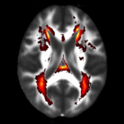
CSVD is typically diagnosed using MRI, which images the brain's white matter. In patients with CSVD, small blood vessels in the white matter become damaged, slowing communication between cells in the brain and, therefore, affecting cognition and motor skills. Fully blocked blood cells can lead to stroke.
But many cases of CSVD are missed because of milder symptoms associated with normal aging, the researchers noted. That's where the blood test comes in, according to Hinman. He and his colleagues identified six proteins related to the immune system's inflammatory response, focusing in particular on a molecule called interleukin-18. Their theory was that inflammatory proteins that damage the brain in CSVD could be detected in the bloodstream.
The study measured protein levels in the blood of 167 people, average age 76.4, who had either normal cognition or mild cognitive impairment. Of these participants, 110 also underwent an MRI brain scan; of these 110 individuals, 49 also underwent a diffusion tensor imaging (DTI) exam.
Study participants whose MRI or DTI tests showed signs of CSVD had significantly higher levels of the six blood proteins. In fact, if a patient had higher-than-average levels of the six inflammatory proteins, they were twice as likely to have signs of CSVD on an MRI scan and 10% more likely to have early signs of white-matter damage.
The investigators also found that for every CSVD risk factor that a person had -- such as high blood pressure, diabetes, or a previous stroke -- the inflammatory protein levels in their blood were on average twice as high.
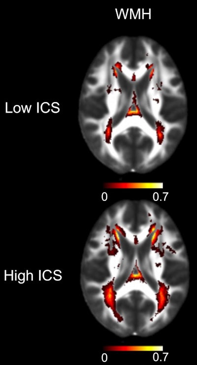 MRI scans showing the average measurable difference in white-matter brain damage in people with low inflammatory blood test scores (below median) and those with high scores (above median). WMH = white-matter hyperintensities; ICS = inflammatory composite score. Image courtesy of UCLA Health.
The blood test could improve CSVD diagnosis because it provides a more quantitative scale for evaluating the disease than does MRI, Hinman said.
"We're hopeful that this will set the field on more quantitative efforts for CSVD so we can better guide therapies and new interventions," he added.


