What's old is new for University of Texas at Dallas (UTD) researchers who are resurrecting an organic, biodegradable compound that someday might be the foundation for a non-gadolinium-based MRI contrast agent, according to a preclinical study published online on February 5 in Chemical Science.
Their focus is on a type of organic radical contrast agent (ORCA) that once was considered for MR imaging, but it was deemed not bright enough for scanning and was broken down too quickly in the body by vitamin C. To correct those issues, UTD researchers have attached these ORCA molecules to a tobacco mosaic virus and encased their creation in a chemical structure to enhance its durability.
"Since this is a plant virus, it can't infect people or animals, and it's easily broken down by the liver. Because the virus is so large, it also allows us to put thousands of the ORCA molecules right next to each other," said co-developer Jeremiah Gassensmith, PhD, associate professor of chemistry and biochemistry at UTD, in a statement. "It's the difference between having one Christmas tree light, which is pretty dim, and a whole string of them together, which is quite bright."
Clinicians certainly are aware of the current concerns over using gadolinium-based contrast agents (GBCAs) for patients undergoing MRI scans. Individuals with poor renal function are susceptible to nephrogenic systemic fibrosis (NSF). While NSF cases have become extremely rare over the last decade, more recent studies have detected elevated signal intensities in brain and liver tissue, as well as bone. This indicates that trace elements of gadolinium still remain in many patients years after their scans.
"Although gadolinium has performed remarkably in clinical settings, concerns about its toxicity, especially for patients with impaired renal functionality, have catalyzed efforts to design alternatives to gadolinium and other metal-based MRI contrast agents," Gassensmith and colleagues wrote.
UTD researchers began the process of developing a new contrast agent by attaching the ORCA to the tobacco mosaic virus. The next step was to create hollow chemical structures, known as cucurbiturils, and wrap them around each ORCA molecule to protect it and ensure that it could maintain its brightness long enough to undergo an MRI scan. This wrapping also allows water to reach the ORCA and tobacco mosaic virus, which is necessary to create the MR image. At the same time, the chemical structure blocks larger molecules from reaching inside the probe and deactivating it or lessening its strength.
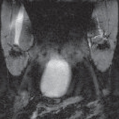
UTD researchers tested their proof-of-concept study on mice and found that the protected version of the ORCA and tobacco mosaic virus provided more than two hours of visible contrast with which to perform preclinical MRI scans on the rodents.
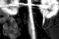
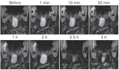 MR images of a mouse before and after an intramuscular injection of the probe show that with the protective molecular structure around the probe, it maintained its contrast for up to two hours. Images courtesy of Gassensmith et al and the University of Texas at Dallas.
"We have some more work to do to show that our material is stable in the complex environment of the human body, and we'd like to see whether we can target it to specific diseases such as cancer and other abnormalities in tissues," Gassensmith noted. "But I think our results are a promising step toward developing ORCAs into clinically viable contrast agents."


