French researchers have identified three key MRI biomarkers to heighten the preoperative detection of a particularly aggressive form of hepatocellular carcinoma (HCC), according to a study published March 31 in Radiology.
The presence of substantial necrosis, high serum alpha-fetoprotein (AFP) levels, and a more advanced stage of liver cancer were independently associated with macrotrabecular-massive HCC, which portends a poor prognosis for patients. Improving the early identification of this HCC subtype could better determine the need for lesion biopsy, more direct treatment, or surgery.
"Pretherapeutic identification of this [HCC] subtype may thus be mandatory for both therapeutic and prognostic purposes," wrote the authors, led by Dr. Sébastien Mulé, PhD, from Henri Mondor University Hospital in Créteil. "Our study demonstrates the ability of preoperative contrast-enhanced MRI to identify HCCs with macrotrabecular-massive subtype."
Hepatocellular carcinoma accounts for the majority of primary liver cancer cases. Macrotrabecular-massive HCC is a serious form of the disease and can result in both macrovascular and microvascular metastases. Contrast-enhanced MRI has shown that it can help in the early and accurate detection of aggressive HCC subtypes, but the modality's contribution to detection of macrotrabecular-massive HCC has not yet been studied.
To help fill that void, Mulé and colleagues performed a retrospective study to analyze 152 patients (median age, 64 years; range, 56-72 years) who preoperatively underwent a liver scan with either 1.5-tesla (Avanto, Siemens Healthineers) or 3-tesla (Skyra, Siemens) MRI. Each patient had one HCC lesion, which subsequently was treated with surgical resection. Among those subjects, MRI identified 26 macrotrabecular-massive HCCs (17%).
To determine which clinical, biologic, and MR imaging features were most associated with macrotrabecular-massive HCC, the researchers looked at a number of variables, which included the following: homogeneity, irregular tumor margins, substantial necrosis, preoperative serum AFP levels, Barcelona Clinic Liver Cancer (BCLC) stage of cancer, and metastases. Three readers then interpreted the MR images, aware of the diagnosis of HCC but blinded to clinical and pathologic findings, including the presence of macrotrabecular-massive HCCs.
They determined that substantial necrosis (occupying at least 20% of the tumor), high serum AFP levels (> 100 ng/mL), and BCLC stage B or C cancer were independent predictors of macrotrabecular-massive HCC subtype:
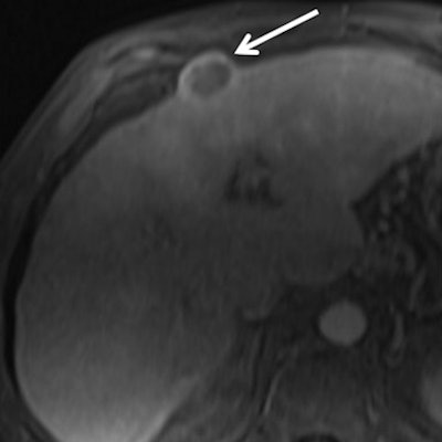
- Through substantial necrosis, 17 macrotrabecular-massive HCCs (65%) were identified, with specificity of 93%.
- High serum AFP level helped identify 13 macrotrabecular-massive HCCs (50%), with specificity of 85%
- BCLC stage B or C cancer helped detect 12 macrotrabecular-massive HCCs (46%), with specificity of 75%.
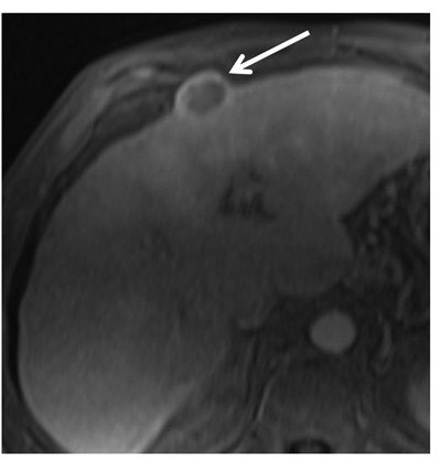
 MR image is from a 60-year-old woman with 2.7-cm macrotrabecular-massive hepatocellular carcinoma (HCC) (arrow). Transverse multiphase MRI scans show the macrotrabecular-massive HCC as a hyperintense central area on T2-weighted image, corresponding to substantial necrosis. Image courtesy of Radiology.
The researchers also analyzed which factors could help determine cancer recurrence within two years or later. In that review, only the presence of metastases was independently associated with both early and overall tumor recurrence.
"Our study highlights the ability of preoperative multiphase contrast-enhanced MRI to identify hepatocellular carcinomas with macrotrabecular-massive subtype with high specificity by demonstrating the presence of substantial necrosis," Mulé and colleagues wrote. "Further investigations using larger multicentric samples of patients are required to validate our findings."


