Imaging findings are illuminating the nature of a newly identified, post-SARS-CoV-2 condition that affects the airways in children called multisystem inflammatory syndrome, according to a study published June 25 in Radiology.
The illness is found in children exposed to or infected with SARS-CoV-2 (confirmed by antibody testing), and causes "airway inflammation and rapid development of pulmonary edema on thoracic imaging, coronary artery aneurysms, and extensive right iliac fossa inflammatory changes on abdominal imaging," wrote a team led by Dr. Shema Hameed of the U.K.'s Evelina London Children's Hospital.
In April, Hameed's group began to see children presenting with symptoms such as fever, headaches, abdominal pain, rash, and conjunctivitis. Clinical findings and laboratory results in these patients showed similarities to Kawasaki disease, Kawasaki disease shock syndrome, or toxic shock syndrome, but were more severe. The team named the condition multisystem inflammatory syndrome in children (MIS-C).
Hameed's team reviewed clinical, laboratory, and imaging findings for 35 children under the age of 17 (median age, 11) who met the criteria for the condition and were admitted to the hospital between April 14 and May 9. Most of the children (94%) had fever; 86% had abdominal pain, vomiting, and diarrhea; 37% had rash; and 26% had conjunctivitis. More than half (60%) were in shock, and 69% were admitted to the intensive care unit (ICU). Of the ICU patients, 20% required mechanical ventilation and 57% inotropic support. Two patients needed extracorporeal membrane oxygenation because of heart dysfunction.
All of the children tested negative for SARS-CoV-2 on reverse transcriptase polymerase chain reaction (RT-PCR), but 30 had serological testing to detect antibodies for the virus; of these, 90% were positive, suggesting previous exposure, the group wrote.
Hameed's group outlined the following imaging findings:
- All 35 patients underwent chest x-ray. Of these, 19 were abnormal, with the most common finding being bronchial wall thickening.
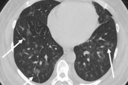
- Thirty-three patients also underwent chest CT because they were suspected for pulmonary embolism due to raised D-dimer and fibrinogen measures. Common findings were basal consolidation and collapsed lung with pleural effusions. No embolisms were discovered.
- Thirty children underwent cardiac CT, which showed abnormal findings such as deteriorating heart function, inflammation of the heart wall and the entire heart, fluid buildup, and coronary artery aneurysms in 51%.
- Five children had abdominal CT scans to exclude appendicitis; the appendix was normal in all but one. Three of these CT exams showed swollen lymph nodes in the right iliac fossa.
- Nineteen patients (54%) had abdominal ultrasound scans. Common findings included inflammation in the right iliac fossa and free fluid in the pelvis.
- Six children had brain imaging with CT or MRI. All of these studies were normal, except one that showed stroke in a child who had been on life support for 24 hours.
The investigators hope the study will help their colleagues be better prepared to treat children presenting with multisystem inflammatory syndrome after SARS-CoV-2 exposure.
"Awareness of this emerging condition and the expected multi-organ imaging findings will aid radiologists in the assessment of these complex cases," Hameed and colleagues concluded.


