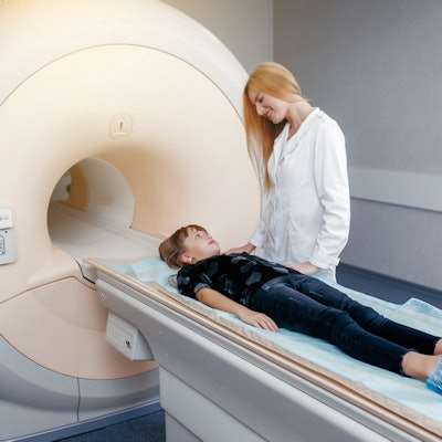
Children present particular challenges when it comes to cardiac MRI, according to a presentation delivered August 9 at the virtual edition of the International Society for Magnetic Resonance in Medicine (ISMRM) meeting.
Dr. John Wood, PhD, of Children's Hospital Los Angeles and the University of Southern California (USC) Keck School of Medicine, outlined a range of technical factors and protocols to keep in mind when imaging this population.
"There are a number of physiological differences between adults and children [regarding imaging, including] vital signs, body habitus, and ability to comply with aspects of the exam [like breath-holding]," he said. "Many children younger than 8 require sedation. And some of our smaller patients have heart and respiratory rates three times higher than adults, which creates some technical challenges [on cardiac MRI]."
Technical considerations
For cardiac MRI in children, 1.5 tesla is a better all-around field strength than 3 tesla -- even though the latter is excellent for cardiovascular indications in adults, Wood said.
"Because of the small field-of-view in cardiac MR for children and the demand for high-resolution images, it's critical that the technician find the smallest coil that fits the anatomy relevant to the study being performed," he noted.
Higher heart rates in children compared with adults can cause blurring on the image, which can be mitigated by lowering the views per segment, Wood said. And children's ability to hold their breath during the exam may be limited.
"We've found that using multiple signal averages is a good [way to control for breath-holding]," he said. "Some children who require an exam with submillimeter resolution may need to be intubated and paralyzed for it."
Different indications
The indications for cardiac MRI differ between adults and children, with adult exams prompted mostly by conditions such as ischemic heart disease or valvular insufficiency, and pediatric exams often indicated by congenital heart disease and iron overload. In his presentation, Wood described pediatric cardiac MRI protocols for a variety of conditions.
Tetralogy of Fallot, a constellation of four congenital defects. The protocol for this indication includes the following:
- Axial cine steady-state free precession (SSFP) whole chest
- Black-blood imaging
- Main, right, and left pulmonary artery flow
- Left atrium/four-chamber SSFP sequences
- Short-axis SSFP stack
- Aorta flow
- Atrioventricular valve, inferior vena cava flow
- Contrast-enhanced MR angiography
- 3D whole heart
Iron overload, caused by blood transfusions necessary for conditions such as sickle cell disease, thalassemia, cancer, or bone marrow transplant. The pediatric cardiac MRI protocol for this indication includes the following:
- Cardiac localizers
- Cardiac T2*
- Cardiac T1
- Cardiac short-axis function
- Aortic flow
- Atrioventricular valve flow
- Liver T2* (three slices)
- Pancreas T2* (15 slices)
- Limited anatomic scanning
Heart muscle imaging, prompted by conditions such as dilated cardiomyopathy or hypertrophic cardiomyopathy. The protocol for heart muscle imaging in children includes the following:
- Axial cine SSFP
- Long-axis, four-chamber, short-axis SSFP cine
- Mitral valve, aortal flow
- T1-weighted, T2-weighted short-axis black-blood imaging
- Quantitative T1, T2 short axis
- 3D SSFP whole heart
- Delayed-enhancement short axis, four channel
- Postcontrast T1-weighted and quantitative T1
Fontan, a surgical procedure for children with univentricular hearts. The protocol for this indication includes the following:
- Axial cine SSFP whole chest
- Black-blood imaging
- Sagittal and coronal SSFP Fontan/Glenn
- Right and left pulmonary artery, Fontan, superior vena cava flow
- Short-axis SSFP stack
- Long-axis, four-chamber SSFP
- Aortic flow
- Atrioventricular valve flow
- Contrast-enhanced MR angiography
- 3D whole heart
- No-contrast lymphangiogram
Helpful framework
In the end, differences between adult and pediatric cardiac MR imaging can be considered from an indication standpoint, Wood said. But it may also be prudent to consider them using a different framework, he concluded.
"I like to think about the principles behind the scanning," he said. "In adults, cardiac MRI is dominated by the need to characterize tissue, while in children, it is dominated by the need to assess anatomy and hemodynamics."


