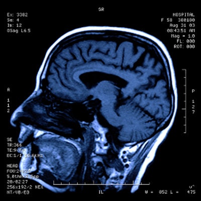
MRI is leading the way in neuroimaging innovation, according to three presentations delivered on Wednesday at the virtual RSNA 2020 meeting.
Neuroradiologists from around the U.S. presented their clinical and research experience with MRI in a variety of novel applications, from using the modality with ultrasound to guide brain disease treatments to diagnosing epilepsy.
MR-guided ultrasound
MRI-guided focused ultrasound (MRgFUS) for brain imaging is showing promise for long-standing clinical problems such as essential tremor, according to Dr. Levi Chazen of Weill Cornell Medicine in New York City. This condition has been treated in a variety of ways since the 1950s, from thalamotomy to stereotactic radiosurgery and deep brain stimulation. Today and into the future, MRgFUS is allowing clinicians to exablate parts of the brain affected by essential tremor.
MRgFUS technology was originally used to treat uterine fibroids and prostate lesions. But over the past 15 years, a cranial device has been developed that focuses a 1,024-element ultrasound array through the calvarium onto a 3-mm focal point; it was approved for treatment of essential tremor by the U.S. Food and Drug Administration (FDA) in 2016.
Patients wear a helmet that transmits the ultrasound to the patient while he or she is inside the MR scanner. The treatment target is the ventral intermediate nucleus (VIM), Chazen said.
"There's a lot of very important and valuable real estate around the VIM that we don't want to ablate, so targeting is really critical for these patients," he said. "With MRgFUS we can precisely tailor our ablation zone to just the anatomy we want to treat."
The procedure is quite effective, according to Chazen.
"As soon as patients come out of the scanner, the tremor is gone," he said. "It's really quite dramatic, and it's not uncommon to see patients in tears."
Future applications for MRgFUS include targeting the internal globus pallidus (GPi) to treat Parkinson's disease and opening the blood-barrier using microbubbles for drug delivery, according to Chazen.
High-field MR neuroimaging
High-field, 7-tesla MRI also shows promise for neuroimaging, according to Dr. Kirk Welker of the Mayo Clinic in Rochester, MN.
"7-tesla MRI has been used in the research realm for years, but in 2017 it received clearance from both the FDA and European Union for clinical use," he said. "Since then, our group has sought to integrate it into clinical MRI practice."
Siting requirements for 7-tesla MRI have become less stringent over the years because the magnets are actively shielded, he said. A larger scanner room is required, but these devices can be installed in an MRI center with 3-tesla and 1.5-tesla systems, allowing for hybrid field strength studies. Currently, the only coils available for 7-tesla scanners are 32-channel head coils and 28-channel knee coils; no body coils have been approved, according to Welker.
The advantages of 7-tesla MRI include increased signal-to-noise ratio -- more than double the ratio of a 3-tesla scanner -- higher spatial resolution (0.2 mm in plane), and improvements in contrast visibility. It also provides increased sensitivity to magnetic susceptibility (that is, how much a material will become magnetized in a magnetic field), which is especially helpful for susceptibility-weighted imaging and functional MRI.
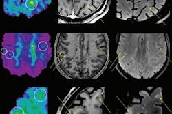
At the Mayo Clinic, Welker and colleagues have found that 7-tesla MRI is particularly helpful for assessing epilepsy because it demonstrates focal cortical dysplasia that can be missed on lower field strengths. It also shows promise for diagnosing mesial temporal sclerosis, another epilepsy-related condition; cavernous malformations; cerebellar hemorrhage; pituitary disease; and multiple sclerosis, as well as being used for presurgical mapping with functional MRI.
Welker did acknowledge that 7-tesla MRI has some significant challenges with artifacts, pulse sequences, and radiofrequency (RF) coil limitations.
"Efforts at parallel transmit and RF coil development will be key to wider adoption of clinical 7-tesla MRI," he said. "But if these challenges are addressed, the proliferation of the technology will undoubtedly continue."
Machine-learning image reconstruction
Deep learning is pushing the boundaries of what neuroradiologists can do, according to Dr. Christopher Filippi of Tufts University Medical Center in Boston. Exciting applications of deep learning for imaging, particularly ultrafast MRI, include improving acquisition speeds, reducing artifacts, and improving echo-planar imaging distortion, he said. In the pipeline are algorithms that could reduce scan doses and speed up scans without degradation of signal-to-noise ratio or contrast-to-noise ratio.
"The goal is to lower costs and improve access to healthcare with faster scanning -- which could reduce healthcare inequity along the way," he said.


