

Is MR imaging in patients with retained bullet or shrapnel fragments dangerous? The incidence of adverse outcomes in this population is low, but a thorough safety assessment is needed, according to a study published August 13 in the Journal of the American College of Radiology.
The findings underscore the need to focus on patient monitoring during MR imaging in order to "detect symptoms early and terminate scanning to minimize chance of injury," wrote a group of investigators led by Paul Armenta, MD, of the Montefiore Medical Center in Bronx, NY.
Individuals with retained bullet and/or shrapnel fragments often undergo MRI, and few studies have reported MRI-related adverse events in patients with these fragments, the group noted. Although the 2023 American College of Radiology (ACR) recommendations suggest caution when scanning patients with retained fragments, they offer limited safety guidance to MRI personnel, it wrote.
"Patients reporting prior gunshot or shrapnel injury at the time of MR safety screening present a particular challenge due to uncertainty regarding foreign body composition, location, and lack of guidance on whether MRI can proceed safely in a specific scenario," the team explained.
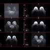
Armenta and colleagues assessed the incidence of retained bullet/shrapnel fragments in patients undergoing MRI and tracked any associated adverse events via a study that included 220 patients with evidence of a retained bullet or shrapnel fragment who underwent x-ray imaging that showed retained bullet or shrapnel fragments and who then underwent a subsequent MRI exam between the years 2010 and 2023. These patients were identified from 6,143 positive x-rays, the team wrote.
The group implemented a safety policy that consisted of assessing patients who reported prior bullet or shrapnel injury or who have evidence of a bullet or shrapnel fragment on review of prior imaging studies at the time of MRI safety screening to locate the fragment (within a solid organ or within 5 mm of a branch of the aorta/vena cava), noting that "these criteria were chosen to minimize potential thermal injury or mechanical force harm to critical structures."
Most of the fragments were identified in the extremities (53.6%), the chest wall (20%), or the abdomen/pelvis (16.4%).
The investigators found that only four of 220 patients (1.8%) reported "transient localized burning or discomfort" during the MRI exam; two of these exams were terminated early and revisited at a lower field strength. In each of these four patients, the metal fragments were in superficial soft tissues (3 in the upper torso and 1 in the thigh). No medical treatment was indicated, and none of the four patients experienced serious adverse events or persistent symptoms.
"In our practice, having a specific institutional screening protocol in place for ballistic fragments has been effective in maintaining and monitoring a safe and consistent MRI service," the authors concluded.
The complete study can be found here.
.fFmgij6Hin.png?auto=compress%2Cformat&fit=crop&h=100&q=70&w=100)


.fFmgij6Hin.png?auto=compress%2Cformat&fit=crop&h=167&q=70&w=250)


