Breast cancer screening with an abbreviated MRI protocol sustains positive patient outcomes over time, according to a study published February 23 in Radiology. The findings could make breast MRI screening more accessible to women.
Breast MRI can be expensive and time-consuming when conducted with a full screening protocol, wrote a team led by Dr. Mi-ri Kwon of Sungkyunkwan University School of Medicine in Seoul, South Korea.
"Despite the high diagnostic performance of breast MRI, its application as a cancer screening tool is hindered by high costs and longer acquisition and interpretation times," the group wrote. "[Abbreviated MRI protocols show] equivalent diagnostic accuracy for cancer while reducing acquisition and interpretation times compared with conventional full-protocol MRI."
Abbreviated breast MRI reduces scanning time from 30 minutes to about 10, the authors noted. Yet despite the promise it shows for breast cancer screening, data regarding the technique's long-term performance have been limited.
Therefore, the group sought to assess the diagnostic performance of a short MRI breast cancer screening protocol over three consecutive years via a study that included 1,975 women who underwent 3,037 exams between September 2015 and August 2018. Of these 1,975 women, 14% were at high risk for breast cancer, 82.7% at intermediate risk, and 3.3% at average risk (this last group had breast MRI by either patient request or surgeon recommendation). The authors tracked cancer detection rate, sensitivity, specificity, positive predictive value, abnormal interpretation rate, and interval cancer rates.
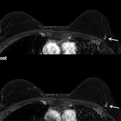
Over the study period, 38 cancers were found; abbreviated screening breast MRI exams identified 29 cancers and missed nine. Of these nine, seven were detected on other imaging modalities and two were interval cancers, for an interval cancer rate of 0.66 per 1,000 exams. All of the nine missed lesions were node-negative, early-stage invasive cancers, according to the group.
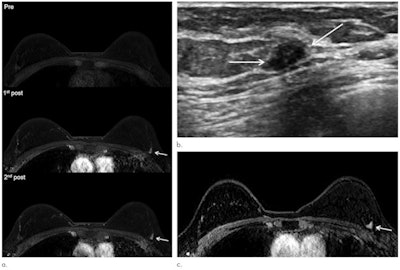
 Representative images of cancer missed at MRI in a 46-year-old woman with a personal history of breast cancer. (a) Retrospectively reviewed abbreviated breast MRI (axial precontrast, first and second postcontrast images) shows a 0.7-cm mass (arrows) in the periphery of the left breast at the 2-o'clock position. At that time, a radiologist mistook the mass as normal background parenchymal enhancement, and it was assessed as Breast Imaging Reporting and Data System (BI-RADS) category 2 on MRI report. (b) After eight months, screening ultrasound image shows 1-cm irregular hypoechoic mass (arrows). Ultrasound-guided core-needle biopsy yielded invasive ductal carcinoma. (c) At preoperative staging MRI, which was performed eight months after abbreviated MRI, mass grew to 1-cm irregular enhancing mass (arrow). Subsequent surgery revealed 1.1-cm invasive ductal carcinoma with 2-cm ductal carcinoma in situ that was intermediate grade, hormone receptor-positive, and human epidermal growth factor receptor 2 negative. Images and caption courtesy of the RSNA.
The abbreviated protocol performed consistently over a three-year time frame, the team discovered.
| Abbreviated breast MRI screening performance over three consecutive years | ||||
| Measure | Year 1 | Year 2 | Year 3 | Total Years |
| Cancer detection rate per 1,000 exams | 6.9 | 8.6 | 10.7 | 8.9 |
| Sensitivity | 75% | 69.2% | 80% | 75% |
| Specificity | 93.5% | 93.3% | 94.1% | 93.7% |
| Area under the curve | 0.89 | 0.84 | 0.90 | 0.88 |
| Positive predictive value for recall | 9.7% | 11.5% | 15.6% | 12.4% |
| Positive predictive value for biopsy | 31.6% | 40.9% | 63.2% | 45% |
| Abnormal interpretation rate | 7.1% | 7.5% | 6.9% | 7.2% |
| Biopsy rate | 2.2% | 2.1% | 1.7% | 2% |
| Short-term follow-up rate | 5.6% | 5.7% | 5.7% | 5.7% |
The study findings suggest that abbreviated breast MRI could further establish the modality's place in the cancer screening toolkit, wrote Dr. Amie Lee of the University of California, San Francisco, in an accompanying editorial. Yet more research is needed.
"This study by Kwon and colleagues confirms that the benefits of abbreviated breast MRI are sustained over multiple consecutive rounds of screening," she concluded. "[Yet] the current study highlights the need for continued long-term evaluation of abbreviated breast MRI performance."


