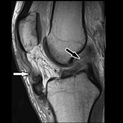
Investigators found that a five-minute MRI protocol for imaging painful knee conditions worked just as well as a 10-minute segment in a study published April 6 in Radiology.
The study results could translate into more efficient, effective patient care -- although more research is needed, wrote a team led by Dr. Filippo Del Grande of Johns Hopkins University School of Medicine in Baltimore.
"[Our study shows that rapid] knee MRI using simultaneous multislice (SMS) and parallel imaging (PI) acceleration can add value through reduced acquisition time but requires validation of clinical efficacy," the group wrote.
Using MRI to diagnose knee conditions is common, and clinicians often choose a parallel imaging acquisition protocol or a simultaneous multislice acquisition to speed exam time. Del Grande and colleagues sought to investigate if the combination of these two techniques could shorten exam time even more, from 10 minutes to five.
The study included 252 adults with painful knee conditions who underwent the following knee MRI protocols between April 2018 and October 2019 on either 1.5-tesla (104 participants) or 3-tesla (148 participants) systems:
- A standard twofold parallel imaging-accelerated 10-minute scan
- A fourfold accelerated scan with parallel imaging and simultaneous multislice acquisition, performed in five minutes
Three radiologists assessed the exams for meniscal, tendinous, ligamentous, and osseocartilaginous conditions; the team compared performance of the protocols at the two magnet strengths using the kappa coefficient.
The researchers found comparable performance across a variety of knee conditions between the shorter protocol and the longer and between the two magnet strengths -- which was supported by a lack of statistical significance between the two protocols.
- Intermethod agreement was good, at greater than 0.71 at 1.5-tesla and greater than 0.85 at 3-tesla (measured by kappa coefficient).
- Diagnostic performance of the two protocols were similar for 1.5-tesla (areas under the receiver operating curve, or AUCs, greater than 0.78) and 3-tesla (AUCs greater than 0.83).
"Our study shows the clinical feasibility of combined SMS and PI acceleration for five-minute, five-sequence ... knee MRI at 1.5-tesla and 3-tesla," the group wrote.
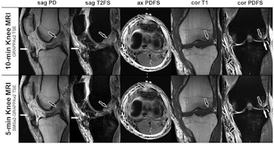 MRI scans in a 28-year-old man with a left knee injury from playing soccer. The corresponding noncontrast axial, sagittal, and coronal MRI scans of the 10-minute parallel imaging-accelerated knee MRI protocol (upper row) and five-minute simultaneous multislice- and parallel-accelerated knee MRI protocol (lower row) were obtained at 1.5-tesla field strength. Sagittal proton density-weighted (sag PD) and sagittal T2-weighted fat-suppressed (sag T2FS) MRI scans demonstrate a tear of the anterior cruciate ligament (black arrows) and a patella tendon tear (white arrows). Axial proton density-weighted fat-suppressed (ax PDFS) MRI scans redemonstrate the patella tendon tear (black arrows) and a tear of the medial collateral ligament (gray arrows). Coronal T1-weighted (cor T1) MRI scans demonstrate a bone contusion of the lateral femoral condyle with small, nondisplaced subchondral fracture (arrows). Coronal proton density-weighted fat-suppressed (cor PDFS) MRI scans redemonstrate the medial collateral ligament tear (short black arrows), a tear of the lateral meniscus (white arrows), and subchondral bone marrow edema pattern (long black arrows). Image courtesy of Radiology.
These findings suggest that rapid knee MRI is within reach, according to a commentary that accompanied the study.
"[This technique is the only one] currently available for clinical use that can reduce acquisition times sufficiently to perform a complete knee examination in five minutes while providing similar diagnostic performance and without reducing the image resolution or compromising image quality," wrote Dr. Naveen Subhas of the Cleveland Clinic in Ohio. "This study brings us one step closer to establishing a new normal: the 5-minute knee MRI."


