Deep learning-accelerated brain MRI improves the detection of acute ischemic lesions compared to conventional MRI because it offers better image quality and significantly decreased exam times, researchers have reported in a study published in Radiology.
The technique could translate to better patient care in that it could improve imaging workflow and cut healthcare costs, wrote a team led by Sebastian Altmann, MD, of University Medical Center Mainz in Germany.
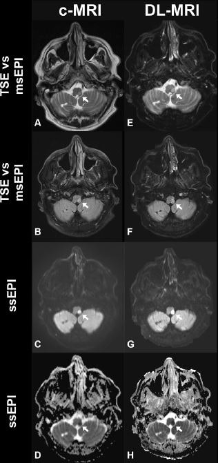
 Example of (A-D) noncontrast transverse plane conventional MRI (c-MRI) and (E-H) deep learning-accelerated MRI scans in a 75-year-old male participant with acute left-sided hemiparesis and a National Institutes of Health Stroke Scale score of 8. Arrows indicate the acute infarction. Furthermore, chronic infarction is depicted in the right biventer lobule. Note the equal image quality. Images and caption courtesy of the RSNA.
"Given the increasing demand for medical examinations and increasing financial constraints placed on health care systems, implementing this technique may be of great value," the group noted.
Deep learning-accelerated MRI can reduce exam times, but research that prospectively evaluates the diagnostic performance of these accelerated MRI reconstructions in acute suspected stroke is scarce, the investigators wrote.
To determine if deep learning-accelerated MRI performed on par with conventional MR imaging, Altmann and colleagues compared the two techniques in a study that included 211 patients who underwent MRI between June 2022 and March 2023. The conventional exam included T1-weighted, T2-weighted, T2*-weighted, T2 fluid-attenuated inversion-recovery, and diffusion-weighted imaging and lasted 14 minutes 18 seconds, while the deep learning-accelerated exam included the same sequences but only lasted three minutes and four seconds (a 78% decrease in exam time).
Three readers assessed the occurrence of stroke, affected vascular territory, overall image quality, and diagnostic confidence; the group set the margin for interchangeability between deep learning-accelerated and conventional MRI at 5%.
Of the 211 study participants, stroke was confirmed in 79. The researchers found that the two MRI techniques were interchangeable when it came to most quality measures and produced high interrater agreement (p > 0.91) -- although deep learning-accelerated MRI showed higher overall image quality and diagnostic confidence (p < 0.001) compared with conventional MRI.
"Unlike traditional acceleration methods, such as compressed sensing, deep learning-based acceleration does not degrade the resolution or appearance of the acquired images," Altmann and colleagues wrote.
The study findings underscore the promise of deep learning-accelerated MR imaging for neuroradiology indications, according to the authors.
"With the ability to acquire images four times faster than the conventional protocol, this technology will speed up the diagnostic process and improve patient care in emergency settings," they concluded.
The complete study can be found here.


