MR elastography (MRE) is an effective technique for noninvasive monitoring of liver stiffness -- a surrogate for fibrosis -- in children and young adults with autoimmune liver disease, researchers have reported.
A team led by first author Jonathan Dillman, MD, of Cincinnati Children’s Hospital Center in Ohio found that MRE liver stiffness measurements were elevated in children and young adults with autoimmune liver disease. The results were published April 18 in the American Journal of Roentgenology.
“[As well, these measurements] were not significantly different between subsets of patients with primary sclerosing cholangitis (PSC)/autoimmune sclerosing cholangitis (ASC) and autoimmune hepatitis (AIH),” Dillman said in a statement released by the AJR.
Liver fibrosis is a key indicator of the progression of autoimmune liver disease, and monitoring it noninvasively would benefit patients, especially young ones, the team noted.
The investigators used data from a registry of children and young adult patients diagnosed with AILD, ASC, or AIH, identifying 46 patients (median age, 16.6 years) who underwent an abdominal 1.5-tesla MRI that included liver MRE as well as a liver biopsy within six months. A 2D gradient-recalled echo sequence for MRE was used to measure mean liver shear stiffness (kPa) for each exam. A pathologist determined histologic METAVIR liver fibrosis stage, blinded to clinical and MRI data (the METAVIR score is a tool used to evaluate the severity of fibrosis; scores range from F0 to F1, normal or mild, to F2 to F4, mild-moderate to moderate-severe).
Median MRE liver stiffness was 2.9 kPa, and MRE liver stiffness measurements correlated with METAVIR histologic fibrosis scores, the group noted. It also reported the following:
| MRE liver stiffness data’s ability to identify advanced histologic liver fibrosis (METAVIR score F2 to F4) | |
|---|---|
| Measure | Finding |
| AUC | 0.81 |
| Sensitivity | 65.4% |
| Specificity | 90% |
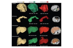
 7-year-old boy with autoimmune sclerosing cholangitis and METAVIR stage 3 fibrosis on liver biopsy. (A) Coronal maximum intensity projection MR cholangiopancreatography (MRCP) image shows multiple intrahepatic strictures (dotted arrows), narrowing of common bile duct (solid arrow), and mild dilation of central biliary tree (dashed arrow). (B) Axial T2-weighted fast spin echo MR image with fat suppression shows enlarged liver with increased T2-weighted signal intensity. (C) Axial MR elastogram image shows heterogeneous appearance of liver with abnormal stiffening (mean liver stiffness, 6.55 kPa). (D) Tissue specimen (Mason trichrome stain; digital scan at 1x magnification) from percutaneous liver biopsy shows abnormally increased fibrosis (blue). Image and caption courtesy of the AJR.
“[Our research showed that] MRE liver stiffness measurements were associated with histologic liver fibrosis severity,” the group concluded. “The findings support a role for MRE in noninvasive monitoring of liver stiffness, a surrogate for fibrosis, in children and young adults with AILD.”
The complete study can be found here.