Multiparametric MRI (mpMRI)-guided targeted biopsy can detect two aggressive prostate cancer subtypes better than systematic biopsy, according to research published July 16 in Radiology.
In a secondary analysis of a prospective trial in patients suspected to have prostate cancer, researchers led by Sangeet Ghai, MD, of the University of Toronto in Canada found that the group of patients who received mpMRI-based targeted biopsy had greater detection of the cribriform (Cr) and intraductal carcinoma (IDC) patterns than a cohort who received transrectal ultrasound-guided systematic biopsy.
CR and IDC prostate cancer subtypes -- an indication of aggressiveness -- are associated with poor clinical outcomes. These subpathologies are “frequently combined for publications as they often occur together, can’t be reliably distinguished on conventional hematoxylin and eosin-stained slides, and have similar prognostic value,” according to the authors.
As evidence for MRI detection of these prostate cancer subtypes has been contradictory, the researchers sought to compare the detection of these morphologic patterns on multiparametric MRI-targeted biopsy with transrectal ultrasound-guided systematic biopsy in biopsy-naïve men at risk for prostate cancer. They performed a secondary analysis of a prospective randomized controlled phase III trial that included patients with a clinical suspicion of prostate cancer at five centers between April 2017 and November 2019.
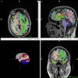
 Multiparametric MRI in a 68-year-old male participant with a prostate-specific antigen level of 9.49 ng/mL. (A) Axial T2-weighted fast spin-echo MRI scan (repetition time msec/echo time msec, 3820/97) shows a 13-mm Prostate Imaging Reporting and Data System 4 nodule in the left midgland posterolateral peripheral zone (arrow). (B) Diffusion-weighted image (b = 1600 sec/mm2) and (C) corresponding apparent diffusion coefficient (ADC) map show marked restricted diffusion at the site (arrows), with an ADC of 515 µm2/sec. (D) Dynamic contrast-enhanced image shows focal early enhancement at the tumor site (arrow). Targeted biopsy revealed grade group 3 prostate cancer (PCa) with cribriform/intraductal histologic features. The participant underwent radiation therapy for treatment of PCa. Images and caption courtesy of Radiology.
Of the 453 participants in the study, 226 were randomized to the systematic biopsy study arm and 227 were placed in the mp-MRI-targeted biopsy arm. In the mpMRI-targeted biopsy group, patients who had a Prostate Imaging Reporting and Data System (PI-RADS) score of at least three received a targeted biopsy.
The investigators reported that 33 (25%) of the 132 biopsies available for assessment in the mpMRI arm identified prostate cancer with the CR or IDC prostate cancer subtypes, compared with 31 of the 196 (16%) biopsies in the systematic biopsy group. The difference was statistically significant (p = 0.01).
In other findings, the researchers observed that Cr/IDC-positive histologic patterns in patients with clinically significant prostate cancer (grade group ≥ 2) had a higher mean cancer core length (11.1 mm) than those with negative Cr/IDC prostate cancer (9.2 mm). The difference was statistically significant (p = 0.009).
“Further studies are required to assess if obtaining more biopsy samples from MRI targets can further increase detection of Cr/IDC [prostate cancer] at biopsy,” the authors concluded.
The full article can be found here.
In an accompanying editorial, Michele Scialpi, MD, of the University of Perugia and Eugenio Martorana, MD, of New Civil Hospital of Sassuolo -- both in Italy -- said that although MRI-targeted biopsy increased detection of Cr/IDC histologic patterns in comparison with systematic biopsy, the results were “not sufficient to confirm its robustness.”
“Further studies are needed to establish the effective role of both MRI and biopsy in diagnosis of the Cr/IDC pattern.” they wrote.


.fFmgij6Hin.png?auto=compress%2Cformat&fit=crop&h=100&q=70&w=100)


.fFmgij6Hin.png?auto=compress%2Cformat&fit=crop&h=167&q=70&w=250)


