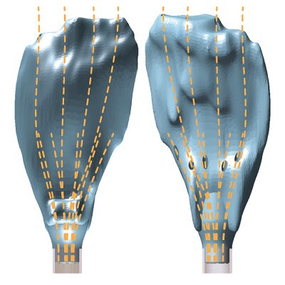
Researchers from the Netherlands have created individually tailored 3D-printed brachytherapy applicators based on the MRI scans of patients with cervical cancer, potentially improving access to tumors during treatment, according to an article published online recently in 3D Printing in Medicine.
Brachytherapy is one of the most widely applied treatments for cervical cancer, and its effectiveness rests largely on the precise delivery of radioactive sources to tumors. The procedure involves using an applicator, a device that contains channels for needles, to guide radiotherapy through either the vaginal or uterine cavity (intracavity) or directly into cancerous tissue (interstitial).
Traditionally, clinicians have relied on commercially available, one-size-fits-all applicators that have a rigid shape, with needle channels at fixed angles. The standardized positioning of the channels in these devices rarely conforms with individual patient anatomy and thus offers a suboptimal distribution of radiotherapy, particularly for large or complex tumors, noted lead author Rianne Laan and colleagues from the Delft University of Technology and Leiden University Medical Center.
To improve upon the safety and accuracy of brachytherapy, the researchers developed a new method for producing a personalized brachytherapy needle applicator using 3D printing technology.
"This approach can offer advantages for reliable applicator positioning within and between fractionated [brachytherapy] treatments, targeting lesions near or behind tissue folds ... minimizing the number of needles required, and enabling proficient treatment for patients with lesions in low-incidence locations," they wrote (3D Print Med, May 16, 2019).
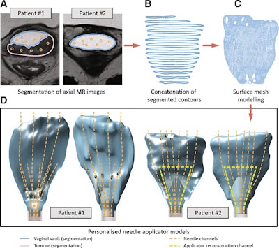
 Workflow for creating 3D-printed brachytherapy applicators based on MRI data. Image courtesy of Laan et al. Licensed under CC BY 4.0.
For their method, Laan and colleagues acquired the pelvic MRI scans of two patients with cervical cancer. Then they used computer software (Oncentra, Elekta) to segment relevant anatomy along with planned needle insertion routes from the scans, which they subsequently converted into virtual 3D models using open-source 3D reconstruction software (3D Slicer). After exporting the models as stereolithography (STL) files, they were able to 3D print the models with a digital light processing printer (Perfactory 4 Mini XL, Envisiontec).
The group tested the utility of the patient-specific 3D-printed applicators on ten gelatin phantoms. An analysis of the tests showed that the applicators yielded effective needle placement for needle channels with a radius of curvature of at least 35 mm. The team also found that the peak force for needle insertion occurred when the target had a depth of approximately 50 mm.
In other terms, a clinician could use these patient-specific 3D-printed applicators and expect highly precise needle positioning for brachytherapy -- with minimal needle jamming or buckling -- as long as the channels meet the minimum radius constraint of 35 mm, according to the authors. The personalization and accuracy that the technique affords may also help bolster treatment planning and even decrease product costs down the line.
"Customized [3D-printed] applicators are expected to stabilize applicator positions, improve lesion access, optimize spatial needle channel distributions, and enhance access to less frequent tumor locations, thereby improving [brachytherapy] treatment conformity, increasing local control in large extensive tumors, and decreasing side effects and their impact on quality of life," they concluded.


