While many reports have praised ultrasound as a useful aid for the diagnosis of appendicitis, researchers continue to examine its potential for greater accuracy in the face of a strong challenge from CT.
Among parameters for evaluating the appendix with ultrasound, Doppler perfusion index below 0.8, round shape, and an anteroposterior outer diameter of more than 6 mm ranked as superior indicators of acute inflammation, according to a series of studies presented at this month's European Congress of Radiology in Vienna.
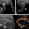
One Austrian study was designed to look specifically at whether the shape of the transverse section of the appendix could be used to include or exclude appendicitis. Dr. Thomas Rettenbacher, from the department of radiology and nuclear medicine at the Hospital Barmherzige Brueder in Salzburg, presented the results at the ECR.
In this study, a total of 382 patients were prospectively evaluated with real-time ultrasound with a 2-4 MHz probe: 174 patients had pain in the right lower quadrant and were suspected of having appendicitis; 108 had acute appendicitis; and 100 healthy patients made up the control group.
In that control group, ultrasound revealed that 37% of the subjects had an ovoid-shaped appendix over the entire length; 30% had a round-shaped appendix over the entire length; and 33% had a combination of both. In the group with a clinical suspicion of acute appendicitis, 37% again had an ovoid-shaped appendix; 24% had a round shape; and 39% had a combination.
Finally, in the cases with acute inflammation, none of the patients had an ovoid-shaped appendix for the entire length of the organ. In contrast, 81% of these patients had a completely round appendix.
"In 25 out of our 175 patients with lower right pain, the ovoid shape over the entire length was helpful to exclude acute appendicitis because other criteria, such as overall diameter or non-compressibility, was misleading," Rettenbacher said. "We can conclude that all acutely inflamed appendixes have a completely or partially round transverse section. An ovoid shape over the entire length reliably rules out acute appendicitis."
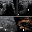
Meanwhile, Italian clinicians assessed vascular changes in acute appendicitis with Doppler ultrasound perfusion and resistive indexes. In this study, 174 patients with suspected appendicitis were prospectively examined with color Doppler. The PI and RI values obtained were compared with those obtained in a 76-person, healthy control group, said Dr. F. Pinto from Cardarelli General Hospital in Naples.
At subsequent surgery, acute appendicitis was found in 162 out of the 174 patients. Of those 162, appendiceal hyperemia was shown with ultrasound in 90.1% of patients. The PI values ranged from 0.3-0.7 in the study population and 0.8-1.4 in the healthy control group. RI values ranged from 0.2-0.4 in the study group and 0.3-0.8 in the healthy contingency.
"There was no overlap in the PI index between the study group and the control group, but there was some in the RI index. PI indexes are more accurate than RI indexes for assessing local vascular change," Pinto concluded.
Rettenbacher questioned Pinto's conclusion on the grounds that "the comparison between these two groups is clinically not so relevant. An analysis of those with acute appendicitis and those with suspected appendicitis would have been more relevant."
Pinto responded that future research would focus on those two areas. This study was intended to serve as baseline data for the range of normal versus pathological values in appendicitis, he said.
Finally, Hungarian clinicians evaluated the diagnostic accuracy of all reported sonographic criteria for appendicitis. Dr. Zolot Tarjan from the Semmelweis Orvostudomanyi Egyetem in Budapest studied the following aspects of the appendix in 178 patients: visibility, anteroposterior outer diameter during compression, maximal wall thickness, the presence of color Doppler signs such as hyperemia, presence of air in the lumen, and presence of inflamed fat. The information was compared to histology in all operated cases.
The appendix was visualized in 86.5% of cases. Using an outer compression diameter of greater than 6 mm as the sonographic criterion resulted in some false-positive diagnoses and unnecessary surgery (sensitivity of 100%, specificity 91.3%), Tarjan reported. If maximal wall thickness greater than 3 mm was used, the false-positive rate decreased, but so did the sensitivity, to 90%. Color Doppler alone had an 81% sensitivity rate and an 80% specificity rate; combining it with outer diameter or maximal wall thickness also improved specificity, but decreased sensitivity.
Air in the lumen excluded appendicitis only if seen in the entire length of the organ, he reported. The measurement of the inflamed fat around the appendix resulted in a false-positive diagnosis in 47% of cases.
While measuring the outer compression diameter yielded the best results, one serious drawback is that many normal appendixes are larger than 6 mm in diameter, Tarjan said. In other cases, where ultrasound found inflamed fat, the problem was found to be not appendicitis but fibrosis. As a result, diagnosing appendicitis can be problematic because morphological signs are not consistent, he said.
By Shalmali Pal
AuntMinnie.com staff writer
March 23, 2000
Let AuntMinnie.com know what you think about this story.
Copyright © 2000 AuntMinnie.com
