(Ultrasound Review) This article, from the Harvard Medical School in Boston and published in the August 2000 issue of the Journal of Ultrasound in Medicine, describes the sonographic findings and diagnostic accuracy of ultrasound in detecting adenomyosis. Adenomyosis is not uncommon in the older reproductive age group. Patients present with abnormal uterine bleeding, dysmenorrhea, and uterine enlargement.
However, the symptoms are nonspecific enough that less than half the patients with adenomyosis have a correct diagnosis made before undergoing hysterectomy. This condition is characterized by an irregular endomyometrial junction, with endometrial glands and stroma extending into the myometrium.
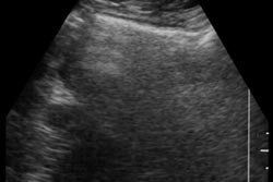
The researchers reviewed the ultrasound findings and hysterectomy reports of 51 women. An ultrasound diagnosis of adenomyosis was made when two or more of the following were present: "a mottled inhomogeneous myometrial texture, globular-appearing uterus, small cystic spaces within the myometrium, and a 'shaggy' indistinct endometrial stripe."
Transabdominal ultrasound was performed using a 4 MHz or 3.5 MHz transducer, and transvaginal examination was added when necessary using a 5 MHz or 5–8 MHz vector-array transducer. Color Doppler was not employed.
Hysterectomy confirmed the ultrasound diagnosis of adenomyosis in 84.3% of patients. Of those patients mistakenly thought to have adenomyosis on ultrasound but who didn’t, 75% had multiple small fibroids, one had stage IV endometriosis, and one case was normal.
The uterus was heterogenous in all patients with adenomyosis, 95% had a globular (rounded) uterus, small cystic areas in the myometrium were shown in 82%, and 82% had a "shaggy" indistinct endometrial stripe. Fibroids were found in the uterine specimen in 63% of these patients, and 33% had endometriosis.
"Although the clinical symptoms of adenomyosis remain nonspecific, the sonographic features of a mottled heterogeneous-appearing myometrium, a globular asymmetric uterus, small, myometrial lucent areas, and an indistinct endometrial stripe should suggest this condition," the Harvard researchers wrote.
J Ultrasound Med 2000; 19:529–534
Bryann Bromley et al
Diagnostic Ultrasound Associates, 333 Longwood Avenue, Suite 400, Boston, MA 02115, USA
By Ultrasound Review
October 17, 2000
Copyright © 2000 AuntMinnie.com


