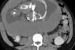
(Ultrasound Review) To assess the effectiveness of pulse-inversion harmonic imaging (PIHI) in detecting acute pyelonephritis (APN), 30 patients along with 10 volunteers were studied using PIHI, tissue harmonic imaging (THI), and conventional ultrasound. The findings of this study, researched by radiologists in Korea, were recently published in the Journal of Ultrasound in Medicine.
The 30 patients with laboratory-proven APN presented with symptoms of upper urinary-tract infection, including fever, chill, and flank pain. PIHI enables improved contrast and spatial resolution and reduces the artifacts generated within difficult-to-image patients. This new technique "generates a pure harmonic signal by setting off the fundamental signals." APN causes focal parenchymal changes, with accompanying blood-flow alteration, and is not reliably demonstrated with conventional ultrasound.
Both kidneys were imaged in longitudinal and transverse with conventional ultrasound, THI, PIHI, and power Doppler using a 2–5MHz curved-array transducer, and patients then proceeded to CT.
Positive ultrasound findings included hypoechoic lesions extending from the renal medulla to the renal capsule, with or without focal contour bulging. APN causes edema, tubular obstruction, and vasoconstriction, so lesions are often triangular or wedge-shaped. Power Doppler findings for APN were simply a focal defect in blood flow.
An analysis of the four sonographic techniques showed a sensitivity and specificity for conventional ultrasound of 57% and 80%, THI -- 97% and 80%, and PIHI with or without contrast -- 90% and 80%. Power Doppler demonstrated a sensitivity of 23% and specificity of 100%, while for CT the sensitivity was 77% and specificity was 100%. The authors suggest that the sensitivity for power Doppler was poor because of technical difficulties such as flash artifacts and large body habitus.
The conspicuity of lesions was graded for the four sonographic techniques. Results demonstrated that both THI and PIHI had better conspicuity than conventional ultrasound, but there was no significant difference between THI and PIHI. In five patients, parenchymal defects were diagnosed using THI and PIHI with and without contrast, but did not show on CT. All four ultrasound techniques incorrectly diagnosed APN-type lesions in two volunteers.
This research is limited by a number of factors that are outlined by the authors, including a lack of bacterial proof in more than half the patients and subjective inclusion and sonographic criteria. "THI and PIHI are sensitive techniques for depicting renal parenchymal lesions and abscess formation in patients with APN," the authors said. "Our preliminary study indicates that these techniques can depict parenchymal lesions better than conventional ultrasound."
"Detection of parenchymal abnormalities in acute pyelonephritis by pulse-inversion harmonic imaging with or without microbubble ultrasonographic contrast agent"
Hyo Keun Kim et al
Dept of Radiology, Samsung Medical Center, Sungkyunkwan University School of Medicine, 50 Ilwon-dong, Kangnam-ku, Seoul 135-710, Korea
J Ultrasound Med (January) 2001; 20:5-14
By Ultrasound Review
March 13, 2001
Click here to post your comments about this story. Please include the headline of the article in your message.
Copyright © 2001 AuntMinnie.com


