Emergency room physicians don't generally perform much ultrasound. They're far more likely to choose radiography or CT for initial imaging exams. A group of emergency department physicians wanted to know why. Among other things, they found that ER doctors needed more ultrasound training, and that radiologists might be more skilled with the modality.
In a study presented at the 2001 RSNA meeting in Chicago, Dr. Max Rosen from Beth Israel Deaconess Medical Center in Boston discussed the barriers that prevent ultrasound from being used more widely in the emergency setting.
"Ultrasound has been used in the emergency department for several years," Rosen said. "It has been used to triage patients, and to provide rapid diagnosis for abdominal and pelvic applications. The purpose of our study was to identify the willingness of emergency department physicians to use a portable ultrasound device for making a diagnosis in the ER, and then to evaluate the performance of the (emergency department) physicians using portable ultrasound."
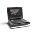
Handheld-ultrasound developer Sonosite of Bothell, WA, contributed five scanner models for use in the study.
Nine emergency department physicians participated in the 12-week trial, which began after the physicians had completed a one-day training session in the use of the devices. The doctors also received additional training in transvaginal ultrasound, on a stationary ultrasound machine equipped with an exam simulator.
The study focused on five specific conditions for which patients were admitted to the emergency room, including:
- Right upper quadrant pain
- Right flank pain
- Blunt abdominal trauma
- Suspected abdominal aortic aneurysm (AAA)
- Vaginal bleeding during pregnancy
"The idea was to identify five indications in which ultrasound could be used to provide a yes-or-no answer," Rosen said.
About 11,400 patients were seen in the ER during the study period. Of these, 1,306 (11%) had indications that did not meet the study criteria, Rosen said. The vast majority of the patients (729) were seen for abdominal pain. As for the rest, 301 had abdominal trauma, 141 had renal colic, and 100 presented with bleeding during pregnancy.
About half of the 1,306 qualifying patients (672) underwent some kind of imaging, either CT or ultrasound. But ultrasound was used only 48 times, Rosen said.
"Again, these were nine attending physicians who were very invested in doing ultrasound, and they had lots of encouragement during the study period, because the emergency department and the co-director of the emergency department were very involved in this," he said.
After 12 weeks, consultants contacted the physicians and interviewed them in order to identify any barriers that might have prevented them from performing more ultrasound exams. The top reasons were:
- A perceived need for additional training.
- A lack of confidence in performing and interpreting ultrasound exams.
- Inability to identify patients who would benefit from ultrasound scanning.
- Discomfort related to the study itself.
- Uncertainty regarding what to tell the patient about ultrasound results, and having to tell patients they would need a second imaging exam for test purposes.
Complete data were available for only 33 of the 48 ultrasound patients, probably due to recordkeeping problems, Rosen said. Compared to CT, the overall sensitivity of ultrasound was 75%, while overall specificity was 89%.
"When you look at the different indications, there were wide variations in sensitivity and specificity," he said. "For patients with right upper quadrant pain, (ultrasound) had 80% sensitivity, 86% specificity. For flank pain, sensitivity was only 40%, specificity was 100%. Blunt abdominal trauma: 50% sensitivity, 67% specificity."
The physicians did very well in evaluating suspected AAA (100%), and also very well in evaluating bleeding during pregnancy (100%), including one instance of fetal demise, he said.
As for specific indications, there were 15 cases of right upper quadrant pain. In 15 patients the gallbladder was not visualized on ultrasound, resulting in one false-positive and one false-negative result, Rosen said.
"In one case when the gallbladder wasn't visualized, the (emergency department physicians) identified liver (metastases), although these were known from prior CT scans," Rosen said. "In the six patients with flank pain, five were positive but only two of five were correctly identified. In one case (ER physicians using ultrasound) missed fibronephrosis, and in 2 cases missed nonobstructing (renal) stones."
Also missed was a large pelvic hematoma. However, the ER physicians correctly diagnosed all but one abdominal blunt trauma patient, and even in that case, free fluid was correctly identified, Rosen said.
He concluded that several barriers prevented the widespread use of ultrasound in the emergency department.
"The issues revolved around a lack of training, and also might have been complicated by the research setting in which ultrasound was performed," Rosen said. "The sensitivity and specificity varied by organ system. In general the specificity was greater than the sensitivity. I think the risk of this is that making the emergency department perform ultrasound may preclude imaging by radiologists, or that findings not related to a simple yes/no paradigm may be missed. The potential (advantages of) ER-performed ultrasound are obviously speed, and potentially triage of patients for more appropriate use of CT or ultrasound performed by radiologists."
By Eric BarnesAuntMinnie.com staff writer
January 3, 2002
Related Reading
Ultrasound disappoints in abdominal trauma, December 14, 2001
Three-year study finds portable abdominal CT useful, November 27, 2001
US, CT replace surgery for some abdominal stab wounds, August 29, 2001
Focused abdominal US in patients with trauma, February 12, 2001
Copyright © 2002 AuntMinnie.com
