(Ultrasound Review) Directed biopsy during contrast-enhanced sonography of the prostate improves the sonographic detection of malignant foci in the prostate. Radiologists and urologists from Thomas Jefferson University in Philadelphia proved this in a study reported in the American Journal of Roentgenology.
According to the authors, both grayscale imaging and color Doppler have proven inadequate in differentiating benign from malignant lesions. "Isoechoic cancers are found in up to 32% of patients with prostate cancer," they said.
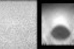
They studied 60 men in order to determine whether "supplemental targeted biopsies using contrast-enhanced harmonic grayscale imaging of the prostate" would improve positive diagnostic yield. Endorectal prostate imaging was performed before ultrasound contrast administration, after continuous contrast infusion and following bolus injection of contrast agent. They used a recently developed contrast agent that consists of liposome encapsulated perfluoropropane microbubbles (Definity, Bristol-Myers Squibb Medical Imaging, North Billerica, MA).
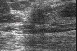
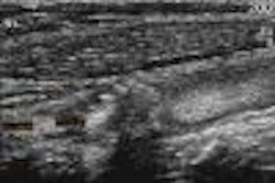
Endorectal ultrasound imaging was performed in transverse and longitudinal using a 6.5 MHz transducer. Precontrast imaging used the fundamental frequency, and scans after contrast administration used wideband harmonic pulse inversion imaging optimized for contrast imaging. An intermittent scanning mode and low mechanical index were used to reduce bubble destruction and lengthen the time the contrast agent was visible.
"Pathology results revealed malignant foci in 30 biopsy sites in 16 of the patients (40%)," they reported. Precontrast imaging showed a sensitivity of 33% and a specificity of 79% for demonstrating cancer. Postcontrast using infusion showed a sensitivity of 40% and a specificity of 81%. The odds ratio for finding cancer went from 2.3 for grayscale to 3.5 for localization of a cancer during contrast enhancement using an infusion method. There was no significant difference in cancer detection between contrast infusion and bolus administration.
They also reported that "a suspicious site evaluated with two cores was five times more likely to have positive biopsy findings than a standard sextant site." Overall 14% of biopsy specimens showed prostatitis, and prostatitis was the cause of false positive findings in 17.6% of cases postcontrast. The authors concluded, "the performance of multiple biopsies of suspicious enhancing foci significantly improves the detection of cancer."
"Directed biopsy during contrast-enhanced sonography of the prostate"E Halpern et al
Department of radiology, Jefferson Prostate Diagnostic Center, Thomas Jefferson University, Philadelphia, PA
AJR 2002 May; 178:915-919
By Ultrasound Review
June 27, 2002
Copyright © 2002 AuntMinnie.com


